UCL prize for paper on pioneering biomedical imaging device
6 August 2010
Links:
 ucl.ac.uk/medphys/" target="_self">UCL Medical Physics and Bioengineering
Researchers from UCL Medical Physics & Bioengineering and the UCL Cancer Institute have won an award for a paper on a pioneering medical imaging instrument that they have developed.
Dr Edward Zhang, Jan Laufer, Professor Barbara Pedley and Professor Paul Beard won the 2009 Roberts Prize for the best paper published in Physics in Medicine & Biology last year for their paper on photoacoustic tomography.
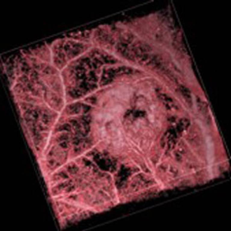
The paper describes a new type of biomedical imaging instrument that exploits the photoacoustic effect - a phenomenon in which the absorption of short laser pulses by haemoglobin can be exploited to produce high-resolution 3D images of blood vessel networks.
The instrument is based upon an optical ultrasound sensor that was invented at UCL. It represents a completely new approach to the implementation of photoacoustic imaging, offering improved sensitivity and resolution over conventional methods.
The study reported in the paper demonstrates that the scanner can provide 3D images of networks of blood vessels in the skin and a tumour model to depths of approximately 5mm with sub-100 micrometre spatial resolution (below a tenth of a millimetre).
The concept offers the prospect of providing a range of improved imaging tools for studying soft tissue abnormalities characterised by changes in the structure, oxygenation and flow status of blood vessels.
Potential applications include studying the development of tumours and their response to treatment, characterising superficial soft tissue damage such as burns or wounds and assessing abnormalities of the microcirculation in patients with lower limb venous disease and diabetes.
Since publication of the paper, a dedicated preclinical version of the scanner has been constructed and recently installed in the UCL Centre for Advanced Biomedical Imaging (CABI).
The scanner is the first of its type worldwide and complements the existing optical, MRI and CT preclinical imaging facilities at CABI. It is currently being used in a range of collaborative studies in cancer, neurology and embryo development.
For more information, follow the links above.
Image: Photoacoustic image of SW1222 colorectal tumour xenograft
UCL context
UCL Medical Physics & Bioengineering is one of the largest medical physics and bioengineering departments in the UK and has very close links to several major teaching hospitals as well as excellent academic research. Research in the department includes medical imaging, physiological monitoring, and the development of implanted devices to identify new technologies and methods for diagnosing, treating and managing diseases.
The UCL Cancer Institute carries out internationally recognised cancer research and promotes the translation of basic research discoveries into new strategies to prevent, diagnose, monitor and cure human cancer.
 Close
Close

