Novel neonatal imaging machine
13 January 2006
A team of scientists from the UCL Biomedical Optics Research Laboratory within UCL Medical Physics & Bioengineering has developed a novel imaging technology that could have major benefits for the diagnosis and treatment of babies in intensive care.

Over the past ten years, the scientists have developed MONSTIR, an instrument designed to exploit a new imaging technique called optical tomography. Optical tomography generates three-dimensional images of biological tissue, using measurements of transmitted light. Dr Adam Gibson said: "Haemoglobin and other natural chromophores absorb visible and near infrared wavelengths, thus allowing the optical tomography technique to reveal information about tissue oxygenation, haemodynamics and metabolism harmlessly, and at the bedside."
One of the major challenges the team faced in the development of MONSTIR was the fact that a beam of light cannot penetrate tissue for more than a few millimetres before it has been completely scattered many times. MONSTIR overcomes this by transmitting very short pulses of light and parallel time- resolved detectors. The device measures the time taken for a photon to travel between pairs of points on the tissue surface. The team have been developing MONSTIR for neonatal brain, breast and muscle scanning.
The data is processed by sophisticated image reconstruction software, called TOAST, developed by the laboratory. TOAST turns the complex optical tomography measurements into three-dimensional images.
MONSTIR has a number of advantages over established methods for imaging the neonatal brain. MRI scans often mean the infant has to be sedated then transported to the machine, which can be dangerous for an infant in intensive care, despite providing detailed information on brain function. Ultrasound scans can be performed at the cot-side and can reveal brain anatomy, but this type of scan cannot reveal brain function.
Dr Gibson said: "The technology we're developing has the potential to produce high-quality images at the cot-side and is also cheaper than MRI. It could make an important contribution to the care and treatment of critically ill babies. Scanners could be available commercially within a few years."
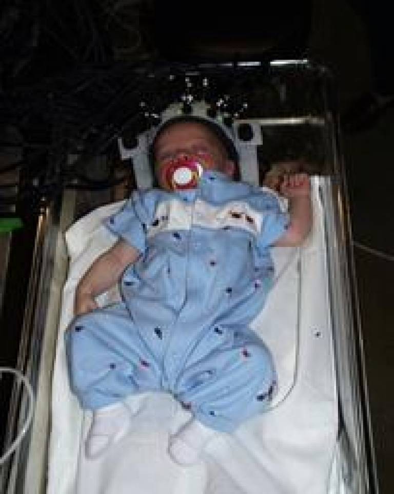
The prototype MONSTIR has been used successfully within the UCLH neonatal intensive care unit since 2001, where it has been used to carry out a number of studies on both ventilated and non-ventilated premature infants. A custom-made helmet containing 32 light sources and 32 detectors fits over the baby's head while still in its cot, and is sufficiently comfortable to be worn for a number of hours.
It takes MONSTIR only eight minutes to generate an image, and the equipment can monitor problems such as intraventricular haemorrhage, changes in blood volume and oxygen, and whole brain imaging of evoked responses.
The machine is about the same size as a fridge-freezer, so the team is now concentrating on producing a new version that is half the size, five times faster and even more accurate.
To find out more, use the link at the top of this article.
Image 1: MONSTIR in use at the cot-side
Image 2: MONSTIR has 32 light sources and detectors
 Close
Close

