 Close
Close

Throughout our undergraduate programmes, students are equipped with the skills needed to address real-world healthcare challenges using the practical application of engineering principles, design concepts and technology.
Here are some examples of the experiential learning you would receive on one of our undergraduate programmes in your first, second and final year:
Second year Biomedical Engineering students participating in scenario week
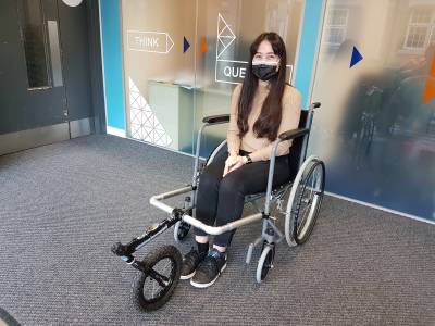
Final year Biomedical Engineering students taking part in the group project; Wheelchair Sports
First Year
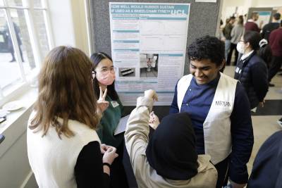
Instron Poster Exhibition
The Instron Poster Exhibition takes place in the first year and is a fantastic opportunity for students to gain valuable experience of presenting research to a panel of expert judges in a conference-style setting.
The expert judging panel comprises of industry practitioners, as well as academics and researchers for UCL Medical Physics and UCL Mechanical Engineering. They are joined by a second panel of more senior Biomedical Engineering undergraduate students who have participated in the same project in previous years and are able to give participating students their unique perspective and advice on the experience.
In participating in the research project, students developed the ability to plan, organise and successfully carry out an experimental study with a strict deadline. They also demonstrated the ability to analyse data using the concepts they learned in class and apply it to a real-world scenario; to think critically; and to compare experimental results with information from other sources.
The winning project, as judged by Instron receives a prize fund and participates in the annual MPBE Student Award Celebration.
“I thought it was really insightful to be able to talk to assessors and academics from different backgrounds as they all had different advice and information to provide. It was also really interesting to talk to them about their career and get an idea of what life is like as a biomedical engineer. The energy at the event was great and felt very casual while still being a formative part of our course. Overall, it was a great experience and I look forward to events like it during the rest of my career.
Ari, MEng Biomedical Engineering, year one
Second Year
Scenario Week
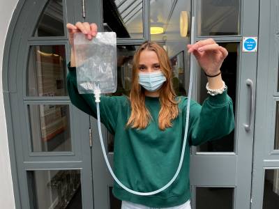
One of the highlights of our Biomedical Engineering degree programme is the scenario week; students are assigned to random groups and tasked with designing a practical solution to a real-world, medical challenge. These are intensive, hands-on design projects that utilise the expertise that students have developed throughout the programme so far.
In 2021-22, the students were set the task of designing and manufacturing a peristaltic pump which maximises V/t of liquid pumped under specific test conditions, while using a closed circuit. In the final demonstration, the teams must set up a closed-circuit and pass liquid using their pump from fluid bag 1 to bag 2. The winning team successfully transfer the highest volume of liquid from bag 1 to bag 2 which has to be leakproof.
Past scenarios have included writing a smartphone healthcare app, developing assistive technology, and creating an article of smart clothing for athletes.
Final Year
Design Project
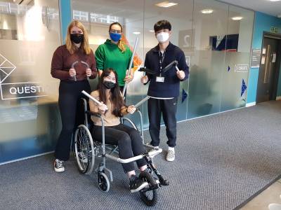
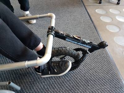
As part of the Design Project, our Biomedical Engineering students gain the experience of creating and building a device that solves a real-life problem for someone with a disability.
Each group works on a Biomedical Engineering related product for a specific user community, e.g. wheelchair users, MND patients, the visually impaired, etc. All the stages of the design need to be considered and addressed, from the initial research and definition of the need and the brief to the design, prototyping, testing, manufacture for commercialisation, addressing IP and regulatory matters, and planning of a route to market.
In the image, we see some students at the prototyping and testing stage, where they discuss what slight modifications they need to incorporate in their design to make it more functional.
Examples of final-year student projects
Here are some examples of past projects that have been firstly outlined by our academics, who then supervised our undergraduate students in implementing those, as their final year projects:
- Mechanical Design of a Brain Implant
We are developing a brain implant to treat epilepsy. The implant uses LEDs which will be bonded to their connection pads. The device will then be encapsulated in silicone polymer which is intended to prevent leakage currents flowing in parallel to the LED due to liquid from the body. The polymer must remain adherent to the surfaces so that there are no voids into which water can condense. If there is too much stress in the polymer after curing, the adhesion may fail, voids will form, and failure will occur. This type of failure is particularly likely underneath the LEDs. To develop our design, we can use a COMSOL model but without some validation, we would not be confident that the COMSOL model is realistic.
This project is to create a scaled-up physical model which will validate the COMSOL model and we hope will give confidence that COMSOL is realistic at real scale. The scaled-up physical model will be made from alumina diaphragms, on which a strain gauge Wheatstone bridge has been formed from thick-film resistor paste. The diaphragms will be calibrated by varying differential gas pressure. They will then be fixed to the ends of short lengths of a glass tube, have some silicone poured on top and cured. Many of these will be made. We want to know how the mean direct stress (= pressure) after curing depends on the depth of rubber, cure temperature, and modulus of the rubber. When these empirical relationships are known, they can be compared with results from COMSOL.
- Experimental evaluation of an optical method for detecting stroke in newborn infants
Optical techniques are being developed at UCL as a means of detecting evidence of stroke in the newborn infant brain. Stroke is caused by blockage of one of the main arteries in the brain and can result in lifelong disability. It usually occurs around the time of birth, with an incidence of about 1 in every 2500 infants. Currently, it is difficult to make a diagnosis of stroke within hours of the event, which is necessary in order to develop treatments to minimise the occurrence of brain injury. Diffuse optical tomography (DOT) involves shining near-infrared (NIR) onto the scalp, which can penetrate several centimetres into the brain where it is strongly scattered, and absorbed by the blood. Light scattered back to the surface carries information about the distribution of blood in the brain and its oxygenation. The aim of this project is to investigate whether measurements of photon flight-times in the head can be used to detect a stroke. Initially, the student will construct a so-called “phantom” – a resin block which has optical properties matched to those of the infant's brain, and which contains regions which simulate the presence of a stroke. The project will then involve performing measurements on the phantom using a sophisticated DOT system, and then evaluate various methods of detecting the stroke regions.
- Implementation of a 3D Magnetic Tracking System for Spatial Registration of Neuroimaging Devices
Registration of the location of the sensors used by non-invasive neuroimaging methods (such as EEG, fNIRS) to appropriate models of the head and brain anatomy is critical to the production of accurate images using these techniques.
The advent of wearable electronic sensors for neuroimaging opens a new path to optimize and automate this registration process. Motion sensors have the potential to allow these neuroimaging devices to provide intrinsic registration of their 3D location in real time.
In this project, the student will be responsible for designing and implementing a proof-of-concept magnetic tracking system for registration of wearable neuroimaging sensors. The student will gain knowledge in fields including neuroimaging, magnetic field modelling, and optical electronics and inverse problems. They will have the opportunity to develop useful skills including programming (Arduino, Matlab), the use of microcontrollers and circuit design. They will also gain significant practical experimental experience. These skills will be applicable in a wide range of future studies and careers.
Approximate Project Outline:
Review literature of magnetic tracking methods, pulsed DC tracking, magnetometers (W1-W2) Familiarize yourself with data output of Invensense MPU chip, magnetometer data sheets and performance (W1-W2) Produce set-up to acquire magnetometer data in real time using development boards (W2-W4) and programmable magnetic field generator Using 3D printing methods, design and develop measurement jig to allow precise positioning of magnetometer relative to field generator (W4-W8) Create detail dataset that encapsulates the measurements obtained from the magnetometer in the field as a function of field generator settings, driving current, position in jig, time, board, etc. (W8-W12) Use this dataset to investigate inverse solutions that yield position in the field given knowledge of the field and measurements taken from the magnetometer (W12-W16) Integrate these solutions to create a prototype, real-time, 3D positioning system
More project examples can be found below:
- Automated segmentation of organs-at-risk for paediatric radiotherapy
Currently, 40-50% of children with cancer receive radiotherapy (RT) as part of their treatment. In RT treatments, the tumour and organs-at-risk (OARs) are manually delineated on planning CT scans, and then the treatment beams are designed to maximise dose to tumour while minimising dose to healthy tissues. Children are particularly sensitive to radiation effects, and so it is very important to quantify and minimise dose delivered to OARs, even if they are not directly in the beam path. However, in clinical practice such delineations are not always available since manual delineation is laborious and time-consuming – automated techniques are therefore highly desirable.
This project will suit an intercalated medical student with interests in radiation oncology and willing to learn computing skills such as MATLAB. The student will create a database of CT images and segmentation of organs at risk in medulloblastoma patients, a common type of childhood cancer. These will be used to test different strategies for automatic organ segmentation, and identify the most promising approaches.
- Development of a dynamic phantom for diffuse optical imaging based on smart film technology
The UCL Biomedical Optics Research Laboratory (BORL) has extensive expertise in the development of physical models of human tissue (known as “phantoms”) which have optical properties matched to those of real human organs. These are used to evaluate new optical techniques and instruments for diagnostic monitoring and imaging, and of the brain in particular. For example, optical imaging techniques are commonly used to observe localised changes occurring in the brain, due to variation in blood flow and oxygenation. The objective of this project is to develop a “dynamic” phantom which can mimic a rapid change in localised optical properties within the brain. The change will be produced by a layer of “smart film” whose optical transparency changes when an electrical voltage is applied. The student will experiment with samples of smart film, and then develop a means of activating the smart film when embedded within a solid block of polyester resin. Pigments will be added to the resin to mimic the scattering and absorbing properties of biological tissues. Finally, images of the dynamic phantom will be generated using the UCL diffuse optical topography system, and its optical properties will be fully characterised.
- Glass-glass sealing for implantable electronics
Implanted electronic devices must be protected from the corrosive environment of the body. One way of protecting electronics is to create a hermetic, or water-impermeable, package which keeps the contents dry. These packages are often made from metals, ceramics, or glasses. You will investigate methods of creating very small hermetic packages by fusing glass layers using high-temperature or laser sintering. A successful student will have knowledge of electronics manufacturing and the attention to detail needed for working in a cleanroom environment.
- Indirect x-ray detectors for medical imaging: comparison between commercial flat panel and custom-built camera
Indirect conversion detectors are widely used in X-ray imaging which is nowadays a key element in many diverse applications, ranging from industrial non-destructive testing to the medical and biomedical fields.
This project involves a complete characterization of a commercial detector based on this technology. It will include classical parameters such as noise and spatial resolution, but also more sophisticated and method-specific ones like signal spill-out between adjacent pixels and detector response as a function of energy, modulation transfer functions and detective quantum efficiency. Subsequently, an X-ray camera will be custom-built by the student, which will entail conceptual design and integrations of the various components. Once the custom camera is built and fine-tuned, it will be characterised by means of the above mentioned quantitative metrics and directly compared to the state-of-the-art commercial solution.
This project is an excellent opportunity to learn the fundamentals of detectors and image analysis as well as for direct hands-on experience in the design and realisation of a custom X-ray camera.
- Detecting anaemia in pregnant women using smartphone photography
Anaemia is a condition in which the haemoglobin concentration in the blood is reduced, impairing its capacity to transport oxygen. The World Health Organisation (WHO) estimated that anaemia affects a quarter of the world’s population. Pregnant women are especially susceptible to developing anaemia. Severe anaemia in pregnant women is also associated with a low birth weight of the baby, stillbirth and newborn death. Once the condition has been identified, most cases of anaemia can be treated effectively. We are developing a smartphone-based technique to diagnose anaemia based on the colour of blood vessels in the eye area. The student will have the opportunity to join our multinational team made up of clinicians and scientists from the UK and India, and participate in the development of a non-invasive imaging technique for screening anaemia in pregnant women. This project will suit a student who is interested in global health, signal and image analysis, statistics and digital photography.
- Development of multimodal imaging tissue-mimicking phantoms for clinical training of minimally invasive procedures
Guidance of minimally invasive procedures can be achieved with a combination of different imaging techniques such as X-ray (computed tomography / fluoroscopy) and ultrasound imaging. Tissue-mimicking phantoms have been shown to be valuable tools for quantifying imaging performance, and for training of young practitioners. This project involves the development and evaluation of new types of multimodal imaging phantoms using open-source software to extract anatomical structures and generate moulds, 3D printing and novel soft tissue-mimicking materials. Potential clinical applications could include liver resection or stent placement. The students will work closely with our clinical collaborators to test and refine these phantoms.
- Improving optical ultrasound generators via surface morphology changes
Optical acoustic generators, where ultrasound is generated using light through the photoacoustic effect, have recently been shown to be a viable alternative to conventional piezoelectric technology. Efficient optical acoustic generators typically consist of a highly optically absorbing dye, which converts incident light into heat, and an elastomeric component that efficiently converts this heat into a pressure increase; typically, polydimethylsiloxane (PDMS) is used due to its high thermal expansion coefficient. In addition, the easy handleability of PDMS allows for a wide range of manufacturing techniques resulting in applications of optical ultrasound that are hard to achieve using piezoelectric technology. However, to this date, the effect of the surface roughness and microstructure on the generated ultrasound and composite coating, has not been investigated.
The aim of this project is to investigate how the surface roughness of the substrate for ultrasound generation affects the generated ultrasound field and the coating uniformity. This work has the potential to change our understanding of how these generators work and to improve future imaging devices. The student will develop these coatings, working with novel nanocomposites and undertake their characterisation, using techniques such as atomic force microscopy. Training in these techniques will be provided. The project will be most experimental in nature; some prior lab experience would hence be advantageous, but not essential.