 Close
Close

Photoacoustic imaging is a new biomedical imaging modality based on the use of laser-generated ultrasound. It is widely viewed as one of the most exciting and promising imaging techniques to have emerged in recent years offering major opportunities for increasing our understanding of biological processes and improving the clinical assessment of cancer and other major diseases. The technique relies upon the absorption of low energy nanosecond pulses of visible or near infrared laser light by specific light absorbing molecules such as haemoglobin to excite broadband ultrasound waves. These waves are encoded with the optical properties of the tissue and, by recording them over the tissue surface using an array of ultrasound receivers, a 3D absorption based image can be reconstructed. The fundamental advantage of photoacoustic imaging is that, by encoding optical absorption on to acoustic waves it avoids the penetration depth or spatial resolution limitations of purely optical imaging techniques that arise from the strong optical scattering of tissue. At the same time it retains the high molecular based contrast and spectral specificity of optical methods enabling visualisation of anatomical features indistinguishable with conventional imaging modalities such as ultrasound. The technique has many potential applications in medicine and biology. Clinical applications include the assessment of breast, skin and colon cancer, cardiovascular disease and dermatological conditions. The technique also has important applications as a research tool in the basic life sciences for studying the biology of a wide range of disease, particularly cancer.
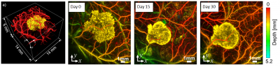
Longitudinal in vivo photoacoustic images of genetically engineered tyrosinase labelled K562 cells in mammalian tissues. Tyrosinase catalyses tyrosine which produces highly absorbing eumelanin and thus strong photoacoustic contrast. The tyrosinase labelled cells (coloured yellow) can therefore be selectively visualised against the vasculature with high contrast at depths well beyond that achievable by in vivo microscopy. (Jathoul AP, Laufer J, Ogunlade O, Treeby B, Cox B, Zhang E, Johnson P, Pizzey AR, Philip B, Marafioti T, Lythgoe MF, Pedley RB, Pule M, Beard PC, 2015, Nature Photonics, 9 (April): 239–46.)
Photoacoustic Imaging Group
The Photoacoustic Imaging Goup was founded in 2003 and forms a sub-group of the UCL Biomedical Optics Research Laboratory, BORL. The group now comprises more than 25 postdoctoral researchers and PhD students making it one of the largest worldwide dedicated to the development of biomedical photoacoustic techniques. The group’s research encompasses the development of photoacoustic detection and excitation devices, non invasive imaging scanners and endoscopic probes, mathematical models of photoacoustic signals, image reconstruction algorithms, spectroscopic methods, and the application of the technique in the clinical and life sciences. For further information, please visit the group website.