Close
Close

Infant anatomical head modelling:
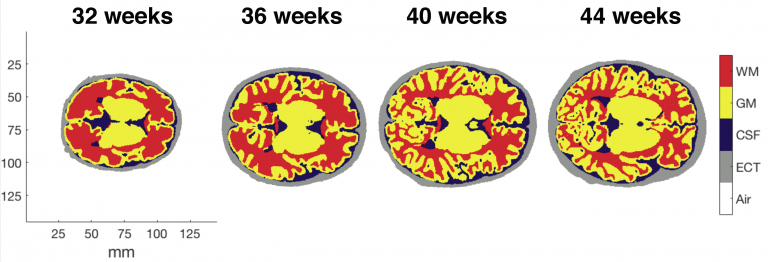
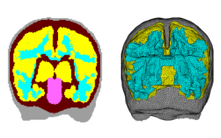
The infant head undergoes rapid structural changes on a timescale of weeks. Diffuse optical tomography (DOT) requires a model of photon transport from sources to detectors through a subject's head - and this requires structural information. A structural prior is a model of the head wherein tissues with different optical properties are delineated. Ideally, a structural prior would be produced using a subject's own MRI scan. However, necessitating an MRI scan for each subject undermines the benefits of DOT, notably its portability and its tolerance of motion that allows the infant brain to be studied in the awake state. At DOT-HUB we are developing anatomical models of the infant head using state-of-the-art MRI data from the Developing Human Connectome Project (dHCP) that can be used when modelling photon propagation within the head. At present we are developing a database of structural for infants aged 28-44 weeks post-conception and quantifying the effect on DOT image quality of using a structural prior from the database as opposed to a subject-specific structural prior.