Close
Close

"With great power comes great responsibility" Pedro, David and Ricardo on their recent publication in Frontiers in Immunology
Pedro Pereira, David Albrecht and Ricardo Henriques tell us about their recent publication in Frontiers in Immunology, where they discuss how fixation protocols can affect super-resolution imaging of the cell cytoskeleton.
What discoveries led you to the research described in your publication?
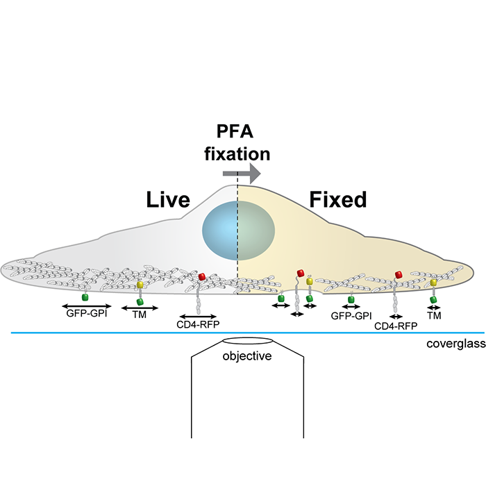
Light microscopy is a key tool in modern cell biology. Conventional light microscopy is limited in its resolving capacity to about ~250 nm, in practical terms this means that any biological details smaller than ~250 nm are blurred and cannot be accurately discriminated. This limitation is critical if we consider for example that all known viruses capable of infecting human cells have sizes close to or smaller than this resolution limit. Super-resolution microscopy is a family of recently developed light microscopy techniques that can overcome this resolution limit. It holds the potential to resolve cellular structures and viruses at near-molecular scales (~20nm). However, as a new field of study, super-resolution microscopy also has some teething troubles due to the long history of established sample preparation protocols inherited from conventional light microscopy. Protocols that were previously acceptable may be inadequate for super-resolution microscopy with its greater resolving power, as the inaccuracies are no longer masked by the blurred image. In this work we aimed to showcase how one key step in sample preparation, fixation, can have a considerable effect if not done properly.
What were you trying to understand?
Our objective was to understand if common fixation protocols that have different effects on the cell cytoskeleton (in this case actin) influence the organization of a protein (CD4) that is homogenously distributed at the cell surface. We used this simple question as a way of showcasing how protocol reliability and artifact identification are essential aspects of super-resolution microscopy. Further, we use our work to highlight different tools that are now available for each researcher to establish the influence of fixation for their own research question.
Why is this important?
Using a famous pop culture reference “with great power comes great responsibility”. Super-resolution microscopy techniques allow near molecular scale resolution (~ 20 nm), a 10-fold improvement to what can be achieved with diffraction limited microscopes. Importantly, nowadays this is possible on commercial systems that have become commonly available through imaging facilities and which can be used without the need for any user specialised knowledge. The potential to routinely and easily obtain biological details at the single protein level can be hindered by established sample preparation protocols that were developed for light microscopes where any inaccuracies were masked by the diffraction limit. Hence, any handling we do of our sample should be made in a way that preserves its original nature and does not introduce artifacts that could lead to misinterpretation of the data collected.
Can you use an analogy to help me understand your work?
Let us imagine you are on a plane high up in the sky. You look down through the window and all the cities seem more or less the same. You could use very large features like the River Thames to discern that you are actually flying over London, but you have hardly any chance to find your home or your workplace. Super-Resolution microscopy would be the equivalent of giving you binoculars to zoom in. This enables you to identify your street specifically, because now you can actually see individual buildings, the local pub or your corner shop. In this case bad sample preparation would be the equivalent to having the plane window full of condensation. Even with the best binoculars you will not be able to resolve and identify small features and no matter what the city is now distorted.
What questions remain to be asked?
There is no such thing as a magic bullet when it comes to a sample preparation protocol. The protocols are as vast as the samples analysed or the techniques employed. This said it would be extremely interesting to understand the specific effects different sample preparation approaches have on the cellular organization, for example extending the analysis we did here to more fixatives, buffers and temperatures.
Written by Pedro Pereira, David Albrecht and Ricardo Henriques