Whole slide analysis with QuPath
We offer image analysis algorithms customised to individual projects involving brightfield or fluorescence whole slide images. This means that we can tailor each project to harvest data from images according to the customers’ needs. Every image analysis project begins with a meeting with the customer to discuss their requirements and feasibility of the project. In this meeting, we determine the specific regions of interest within the tissue section, cells of interest, and desired data outputs such as cell counts/mm^2, cell A to cell B ratio, or percentage of positive cells to total cells.
At IQPath, we are uniquely positioned to perform image analysis within a fully integrated facility ranging from the processing of specimens (tissue sectioning, staining) to high-throughput slide scanning and storage before performing image analysis. This means that we can deliver high quality work seamlessly at every step of the process, ensuring that the data you receive is representative and meaningful.
Our image analysis service costs £48 per hour.
Please contact us for further information:
Sebastian Brandner s.brandner@ucl.ac.uk
Yau Mun Lim yau.lim@ucl.ac.uk
Legacy projects
Whole slide analysis with Definiens Tissue Studio and Developer (UCL collaborations)
We can analyse brightfield or fluorescence whole slide images with a digital image analysis software, Definiens Tissue Studio and Definiens Developer. This software is ideal for detection and quantification of biomarkers in cells and tissues. We can analyse slides using predefined algorithms in Definines Tissue Studio or we can develop new algorithms using the Definines Developer Software. Please contact us for further information (Sebastian Brandner s.brandner@ucl.ac.uk or Matthew Ellis m.ellis@ucl.ac.uk).
Read our recent publications from our team using Definiens Tissue Studio
Examples of digital image analysis
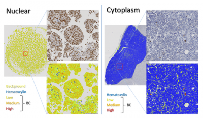
Analysis of experimental brain tumours.
Nuclear or cytoplasmic markers were quantified in a series of experimental brain tumours.
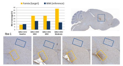
Analysis of microglia upregulation.
Quantification of microglia response in a circumscribed area of mouse brains (IBA-1 immunolabelling).

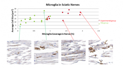
Analysis of intraneural microglia/macrophages.
Comparison of experimental and control samples and correlation between size and frequency.

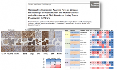
Tumour cell analysis
Selection of regions of interest (left: tissue culture with tumour spheres; right: well demarcated brain tumour) and automated separation of marker-positive from marker-negative tumour cells.

 Close
Close


