 Close
Close

Research at the UCL TouchLab focuses on the development of novel imaging, robotic and computational methods to analyse, explore and manipulate microscale environments.
Researchers in the TouchLab are developing new methods and tools for visualisation and robotic manipulation of biological systems. We work with imaging and computational analysis techniques to extend the capacity of the optical microscope to extract diagnostic information from histological tissue sections and light-driven microrobots for collection of therapeutic targets, such as stem cells, from resected tissues. By collaborating with researchers from across the health and life sciences we aim to deploy these methods to improve clinical and pre-clinical workflows.
Research Highlights
- Fourier Ptychographic Microscope platform
Working with Prof Fernandez-Reyes we have developed a Fourier Ptychographic Microscope platform for diagnostic imaging of blood films, focusing in particular on assessment of cell morphology and the detection of malarial parasites.
- Automated navigation of optoelectronic microbots
Working with the Wheeler Microfluidics Laboratory at the University of Toronto we have developed a method for automated navigation of optoelectronic microrobots which allows simultaneous control of multiple microrobots. Our work is a significant step towards using microrobots for collection of therapeutically important targets from resected tissues.
“Autonomous object harvesting using synchronized optoelectronic microrobots”, Bendkowski et al.
- Light field microscopy of C. elegans
We have applied light field microscopy to explore the functional and behavioural properties of the model organism C.elegans, including its ability to sense mechanical stimuli and the effects of mutations in cuticle collagen genes on locomotion.
YouTube Widget Placeholderhttps://www.youtube.com/watch?v=d_aGM90erYI
Collaboration Case Study
Histopathology, which relies on microscopic examination of excised tissues, remains a cornerstone of medical practice from the diagnosis of disease and assessment of the effectiveness of surgical interventions, to the development and validation of new therapeutic treatments. However, the histopathologist’s ability to visualise and analyse tissues is limited by the capacity of conventional optical imaging systems to sense and capture the full complexity and range of diagnostic information contained within even relatively modest (~mm) sized tissue sections. A reliance on chemical staining reagents increases the workload of the clinical team and the time to diagnosis / assessment, delaying further surgical intervention. Staining variability can also lead to misdiagnosis and renders the tissue unsuitable for analysis using other methods.
Through WEISS we are collaborating with Dr Alex Freeman and his team at UCLH to develop and apply a novel form of computational imaging based on Fourier Ptychographic Microscopy (FPM) to allow efficient high-resolution histological imaging of prostate tissue biopsies. FPM expands the capacity of the optical microscope to capture sample information enabling large format imaging at high spatial resolution for rapid visualisation of large tissue sections. By capturing both amplitude and phase information FPM also allows label-free visualisation of tissue structure, potentially reducing requirements for tissue processing. We believe the method holds particular promise for intraoperative pathology.
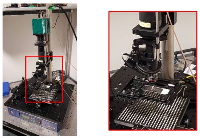
Fourier Ptychographic Microscope (FPM) system in the UCL TouchLab. The LED matrix (rectangular array of white blocks) is used to illuminate the sample at different angles which allows increase of image resolution and recovery of the sample phase.
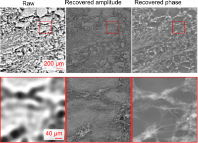
Example raw (left) and reconstructed high resolution FPM amplitude (centre) and phase (right) images of a histological tissue section.