Shedding light on liver surgery with photoacoustics
7 August 2018
A £493,957
 cancerresearchuk.org/">Cancer Research UK grant awarded to Professor Paul Beard, Prof Brian Davidson and colleagues has the potential to boost outcomes for liver cancer surgery. Their research will develop a pioneering new imaging technique, known as photoacoustic imaging (PAI), for use in laparoscopic liver surgery for the first time. The heightened image contrast provided by PAI should make the site and extent of the cancers easier for surgeons to detect and guide their surgical excision.
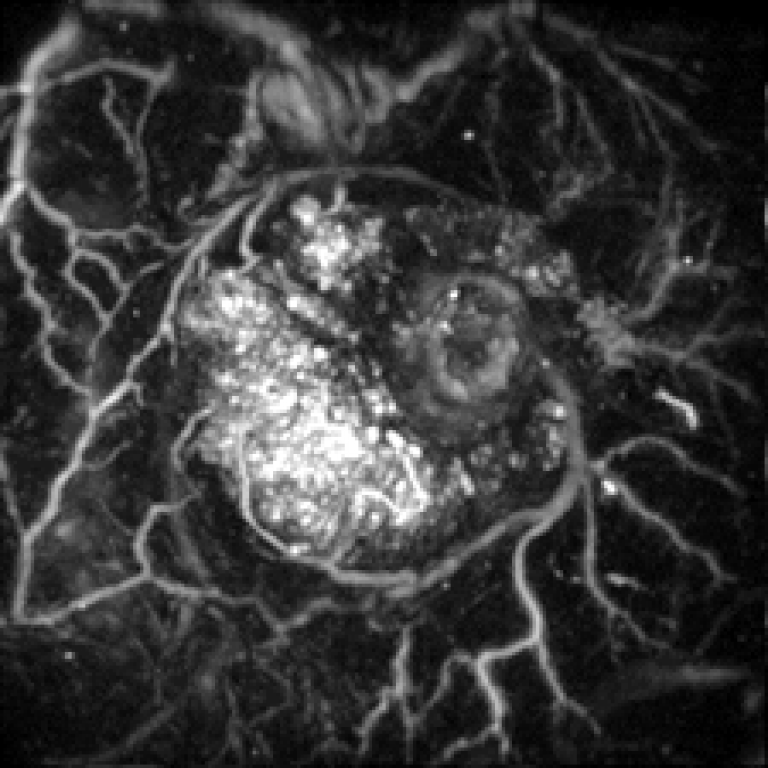
Image of a colorectal tumour, illustrating the capabilities of PAI
Liver cancer affects approximately 1.4 million people globally each year. Surgery is the main form of treatment and around 2,300 resections are performed annually in the UK.
Laparoscopic surgery is a minimally invasive procedure which results in less pain, faster recovery and lower costs than open surgery. However, it is very technically challenging. Due to the small incision, surgeons have limited vision which increases the risk of cancerous tissue going undetected.
To prevent cancer recurring, all affected tissue needs to be removed, while at the same time as much of the healthy liver should be preserved to ensure recovery of liver function and regeneration. This delicate balancing act requires the ability to precisely detect tumours and identify their margins - currently beyond the capabilities of conventional imaging techniques.
Photoacoustic imaging (PAI) is an emerging technique based on laser-generated ultrasound waves. It can provide high-resolution 3D images at centimetre-scale depths. A key advantage of PAI is image contrast is defined by optical absorption. This makes it well-suited for seeing blood vessels and measuring oxygen saturation. The main form of liver cancer, hepatocellular carcinoma, shows a greater density of small blood vessels and higher oxygen saturation than neighbouring healthy liver tissue. As a result, PAI is expected to offer a much clearer view of tumours than conventional ultrasound imaging which cannot reveal tumour vessels with sufficient contrast or measure oxygen saturation.
This is a potentially pioneering development in the treatment of human liver cancer - PAI has never before been applied in this context. By improving the efficacy of laparoscopic liver cancer surgery and decreasing the associated risks, more patients could access this form of treatment. This technique could light the way for the minimally-invasive treatment of other abdominal cancers, such as pancreas and kidney cancer.
"This project would not be possible without a multidisciplinary collaboration between physicists, engineers, surgeons and radiologists," Paul explains.
Professor Paul Beard works within the UCL Department of Medical Physics and Biomedical Engineering, UCL Institute of Healthcare Engineering and Wellcome / EPSRC Centre for Interventional and Surgical Sciences. This project is in close collaboration with clinical lead, Professor Brian Davidson and other colleagues at UCL and Royal Free Hospital.
The newly awarded Cancer Research UK grant will fund this research for 3 years. Cancer Research UK is the world's largest independent cancer research charity. It conducts research into the prevention, diagnosis and treatment of cancer.
 Close
Close

