Research advancement using brain-computer-interfaces may lead to more developed communication with patients unable to move or speak
20 December 2017
Research led by Dr Jinendra Ekanyake using real-time fMRI to decode brain function has recently been published in NeuroImage.
 The results have significant potential applications for using
brain-computer-interfaces (BCI) as a platform for communication in paralysed
patients or those suffering from locked-in-syndrome.
BCIs interpret and use brain activity to control external devices, such as control of a cursor on a screen. Current research has only demonstrated this in primary motor and sensory brain regions, allowing for communication that has mostly been limited to 'yes/no' responses or basic motor functions.
Dr Ekanyake's team's research has potentially opened the path to a more developed degree of communication by providing proof-of-principle for the use of higher-order brain regions, which are involved in more complex cognitive processes such as attention. This is the first time these cognitive, higher-order brain regions have been decoded.
The study examined three brain regions, the parietal lobe, lateral occipital cortex (LOC) and fusiform face area (FFA), which have all been suggested to contain salience maps and have roles in integrating position and category-specific information that allow individuals to 'map out' the world around them. By using real-time fMRI, Dr Ekanyake's team were able to link individuals' brain activity to their covert allocation of attention on real-world images presented at different locations. They were able to reach 70% accuracy in decoding these brain activations in some participants.
'Cognitive BCIs', as demonstrated by this research, represent a significant step forward from the 'yes/no' answers form of communication in their ability to access varied and more versatile information about individuals' mental processes. The clinical applications of this for patients who are unable to speak or move due to an injury or condition are potentially transformative.
Dr Jinendra Ekanayake is an academic neurosurgeon with a research focus on functional neuroimaging, brain computer interfaces and neuromodulation. He has recently been appointed as Senior Clinical Research Fellow to Wellcome EPSRC Centre for Interventional and Surgical Sciences (WEISS). Dr Ekanayake's work highlighted in this paper is a collaboration with UCL Faculty of Brain Sciences and the Wellcome Centre for Human Neuroimaging. The full paper, titled "Real-time decoding of covert attention in higher-order visual areas", is available now online on ScienceDirect.
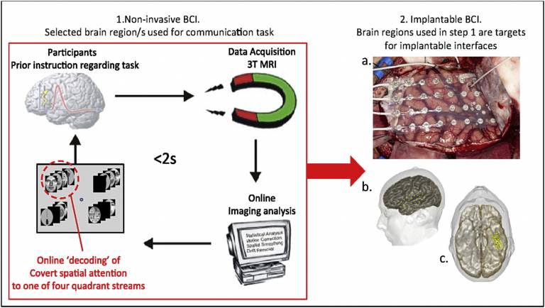
Figure 1: Proposed pipeline using a non-invasive BCI interface with rt- fMRI to prime and prepare specific brain regions with a BCI task, prior to surgery for placement of longer-term implantable BCI. 1) Realtime-fMRI decoding pathway (e.g. as used in this study) 2) A. Implantation of subdural electrodes B, C. 3D reconstruction showing final placement of temporal and inferior temporal subdural (ECoG) grids for recording of relevant cortical activity, as part of a long-term implanted BCI. Thanks to Anna Misserochi for the intraoperative grid placement image.
 Close
Close

