4-Year PhD Studentship: Magnetic Resonance Imaging for non-invasive cancer diagnosis
Now Closed

15 April 2020
Primary Supervisor: Dr David Atkinson (Department of Medicine, Centre for Medical Imaging)
Secondary Supervisor: Dr Eleftheria Panagiotaki (UCL, Centre for Medical Image Computing)
Project summary
This 4-year PhD studentship is a collaboration between Philips Healthcare and UCL. The funding covers an annual tax-free stipend (at least £17009) and tuition fees. As this is a Philips/EPSRC funded studentship the standard EPSRC eligibility criteria apply, please see the EPSRC website for details. The successful candidate will be enrolled onto the UCL CDT in Intelligent Integrated Imaging in Healthcare (i4health) and benefit from the being part of a cohort of PhD students as well as participation in the activities and events organised by the centre.
Background
Magnetic Resonance Imaging (MRI) is now a first-line investigation for suspected prostate cancer, enabling about 28% of patients to avoid a needle biopsy. Nevertheless, in current MRI prostate studies, about 1/3 of cases are classified by radiologists as ‘indeterminate’ and subsequent biopsy results are negative for about half of those performed.
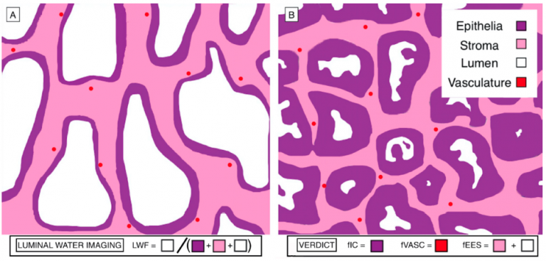
Newer MRI sequences and computational processing techniques that are sensitive to multiple tissue components within each pixel have shown promise for more specific characterisation of prostate cancer. These sequences are already being tested in clinical trials at UCL and include diffusion-based VERDICT and multi-echo T2-based Luminal Water Imaging (LWI).
In breast cancer, false positives from mammography lead to unnecessary stress, cost and biopsies. In the US, the chance of a false positive during 10 years of annual screening exceeds 50%. Mammography also carries a small dose-related risk. Hence there is a desire to improve tissue characterization from MR to provide more accurate diagnostic information. The glandular tissue of the breast has some similarities with prostate microstructure but the diffusion VERDICT and LWI sequences and analyses are relatively unexplored for the breast.
Research Aims
The overall aim is to create new MR sequences and processing techniques that will aid in the management of cancer patients. We hope to improve the diagnostic performance for prostate MRI and investigate application to breast and other cancers.
The project will model the luminal water index acquisition to understand the trade-offs between the accuracy of data fitting, scan duration, image resolution and signal to noise (SNR). A better understanding of acquisition parameters, SNR and their interaction with fitting is likely to ensure a process that is more robust to variations, e.g. patient size, introduction of new coils or simultaneous multi-slice sequences, and enable us to more rigorously set the threshold between malignant and healthy tissue. Alternative fitting strategies, including machine learning, will also be investigated and possibly lead to new, more informative, acquisition strategies.
The diffusion VERDICT and LWI schemes used for the prostate will be adapted for breast imaging.
The student will have the opportunity for hands-on experience with the MR scanner, implementing these cutting-edge sequences for prostate and breast cancer. Evaluation will use subjects recruited from the UCLH clinical imaging pathway, or those part of on-going, funded clinical trials.
Previous work from researchers at UCLH and colleagues has led to changes in national guidelines for the management of suspected prostate cancer patients. The significance of the work in this project is that in partnership with on-going trials, the work has the potential to impact the recommended screening or assessment pathways that will help manage prostate and breast cancer patients.
The project plan has been adapted to cope with the implications of the covid-19 pandemic. This project has data processing and analysis components that can be performed remotely when necessary. We also have scanner simulators that enable remote sequence development. For physical scanner access, a research MR scanner within University College London Hospital (UCLH) is located in an area that is being kept ‘covid-free’ with working protocols to protect researchers, staff and patients. This scanner also has extensive time already funded by a charitable donation to enable access for researchers developing new imaging methods. This time will be available to this PhD student. UCLH is part of an active research environment on prostate and breast cancer MRI with multiple on-going, funded studies.
Requirements
Applicants are expected to have a first degree in Physics, Computer Science or Biomedical Engineering or relevant Physical Sciences based subject passed at 2:1 level (UK system or equivalent) or above. Good working knowledge of computer programming is required. Knowledge of MATLAB and some experience with medical imaging and/or medical image analysis is also desirable.
Deadline 31st July, 2020.
To Apply: Please send a CV and Covering Letter expressing your interest to Dr David Atkinson d.atkinson@ucl.ac.uk
 Close
Close

