 Close
Close

Hermes at UCL
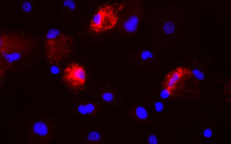
High-content microscopy is an emerging technology which enables single cell analysis of fluorescent staining in large populations of cells, with the ability to investigate subcellular features and localisation. We operate a Hermes imaging system in collaboration with IDEA Bio-Medical in the UCL Cruciform Building, Gower Street, London which allows for automated image capture and analysis of data in a variety of bioassays.
The integrated Athena analysis platform enables simple and rapid quantitation to produce numerical results with minimal training required. For more advanced studies the Minerva analysis package provides the flexibility to meet the needs of most applications. We also feature interesting images each week on our twitter page: @UCLHermes.
How do I use the service?
Experiments and/or training can be arranged by contacting ii.hermes@ucl.ac.uk. A general workflow is as follows:
1. Consultation with the user to discuss the desired assay format and advise on how best to achieve the desired results
2. Users prepare and stain their samples before handing them over to the unit
3. Plates are imaged in the requested channels
4. Image data is computationally analysed to quantify staining characteristics
5. Captured images, overlays and analysis results are handed over to the user.
Users are also welcome to image and analyse their own samples.
Sample applications
- Quantitation of viral transduction
- Detection of bacterial infection
- Multi-colour analysis of protein expression
- Quantification of cellular toxicity
- Nuclear translocation of transcription factors
- Analysis of cell morphology
- Single molecule RNA FISH
- Detection of rare subpopulations within a culture.
 as seen under the Hermes at 4x")