 Close
Close

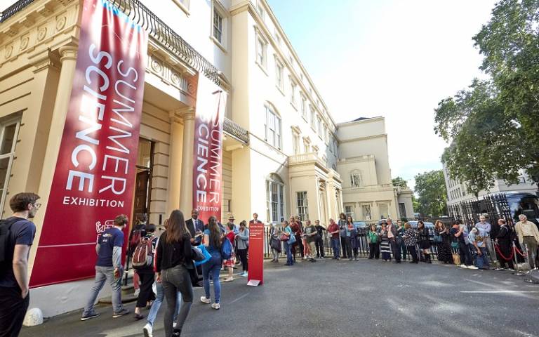
UCL academics exhibit at the Royal Society
10 July 2019
Two UCL teams from the Faculty of Engineering exhibited at the Royal Society Summer Science Exhibition last week.
REANIMATE, from the Institute of Healthcare Engineering, gave the public the opportunity to explore and understand virtual cancerous tumours.
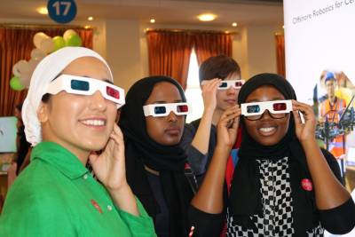
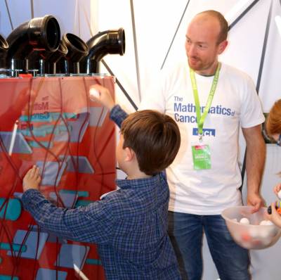
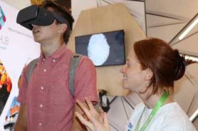
The academics designed their exhibit, The Mathematics of Cancer, to include a virtual reality game allowing attendees to travel through and explore the unique and irregular architecture of a tumour, an arcade-style game demonstrating the difficulty of drug delivery and a game that allows visitors to create virtual tumours using 3D printed tumour ‘slices’.
The team behind the REANIMATE platform (REAlistic Numerical Image-based Modelling of biologicAl Tissue substratEs) have combined advanced imaging techniques with mathematical modelling to gain a better understanding of drug delivery in complicated tumours.
“We are so excited by the opportunity to showcase at the Royal Society and to share our research with the diverse audiences that the exhibition attracts. Not only are we hoping that they leave with a greater understanding of and interest in the ways we are tackling cancer, but also that their questions shine fresh perspectives on our research and shape it moving forward.”
The exhibit demonstrated how the structure of cancer tumours varies widely, making the delivery of therapeutic drugs and predicting their uptake and distribution challenging.
The team also had 3D printed models of different tumours on show to demonstrate their complexity.
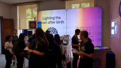
The exhibit, Lighting the Way to a Healthier Brain, included hands-on demonstrations showing which colours of light penetrate the body to reveal blood vessels, plus the chance to image your own brain oxygen and metabolism levels whilst undertaking maths exercises.
The team developed the compact broadband near-infrared spectroscopy (NIRS) system, called CYRIL (Cytochrome-c-oxidase Research Instrument and appLIcation) to diagnose brain injury in newborn babies and enable the implementation of treatment strategies as early as possible.
Dr Ilias Tachtsidis (UCL Medical Physics & Biomedical Engineering) and exhibit leader said: “Doctors already use light sensors to measure tissue oxygen levels, but this gives a limited picture of the brain’s health. CYRIL goes further by using a wider, broadband spectrum of near-infrared light with hundreds of different near-infrared colours to get a much more detailed picture that includes metabolism – how much energy the brain is using.
“Not every hospital has an MRI machine due to the significant costs involved in purchasing and maintaining one. We hope with CYRIL we have a realistic opportunity to actually have a device in every hospital that can give information about the brain oxygenation and metabolic status at the baby’s cotside within a few hours of birth.”
A lack of oxygen and blood flow during birth, also known as Hypoxic Ischemic Encephalopathy (HIE) is a dangerous condition affecting two to three babies out of every 1,000 born in the UK. Cooling treatment helps many to make a good recovery, but nearly half of those affected go on to develop cerebral palsy or cognitive and behavioural problems. It is the second most common cause of preventable childhood disability.
MRI scans can only be done once a newborn is stable enough to be put through an MRI machine, whereas CYRIL can pick up signs of injury minutes after birth, enabling treatment to be started earlier and therefore reducing the likelihood of permanent disability or even death.
CYRIL is also cheaper than MRI scans, which cost the NHS millions of pounds, as the device costs just £50,000 and can be operated easily. It is easily portable in a backpack and UCL academics hope that one day it could be used in lower income countries with limited access to MRI machines.
A version of this article was first published by UCL News on 2 July
Related links:
- Royal Society Summer Science Exhibition
- Dr Paul Sweeney's academic profile
- REANIMATE
- Dr Ilias Tachtsidis’ academic profile
- CYRIL
- UCL Mechanical Engineering
- UCL Medical Physics & Biomedical Engineering
- UCL Engineering