Close
Close

UCL scientists create virtual tumours to aid cancer drug delivery
10 October 2018
Scientists have designed a virtual modelling technique which can create highly detailed 3D models of individual cancerous tumours and simulate the delivery of drugs in order to predict effectiveness.
As featured in today's The i paper, researchers acquired high-resolution images of surgically-resected tumours and used mathematical modelling to run detailed computational experiments. This allowed them to study the transport of blood, biological fluids and drugs, and their complex interactions with tissue.
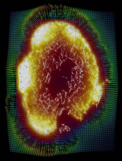
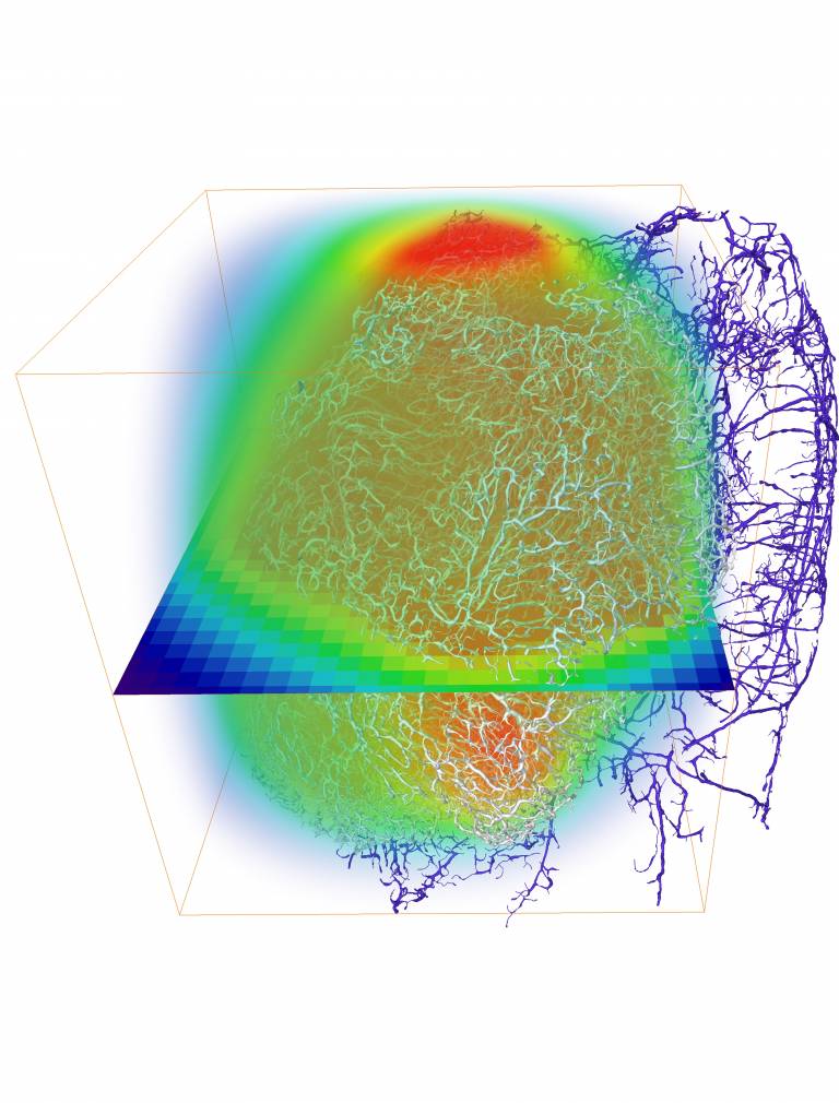
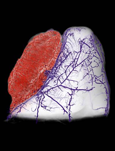
Their new technique, named REANIMATE (REAlistic Numerical Image-based Modelling of biologicAl Tissue substratEs) enables researchers to visualise and interact with large, 3D, virtual models of tumour tissue samples and treat them as living specimens. This will enable scientists to perform complex computational experiments to generate new insights into how individual tumours react to specific treatments.
In the study, published in Nature Biomedical Engineering, researchers used optical imaging of extracted tumour tissue that had been rendered transparent using a cocktail of chemical treatments. These can show fine detail such as blood vessel networks and cell nuclei, which can be seen across entire organs at very high resolution by using fluorescently-labelled probes that bind to specific structures.
Joint lead academic Dr Simon Walker-Samuel (UCL Centre for Advanced Biomedical Imaging) said:
These advances are a truly interdisciplinary effort and would not be possible without the combined input of physicists, mathematicians, cancer biologists, clinicians, imaging specialists and engineers. The new framework has a vast potential impact in helping to develop new cancer drugs and potentially providing a cost-effective way to test their efficacy before going to human trials. It advances the move towards truly personalised medicine, with the potential aim that one day clinicians might be able to predetermine the most effective therapeutic plan for each patient’s unique tumour makeup.“
It is predicted that one in two people in the UK will be diagnosed with cancer in their lifetime, with cancer treatment expected to cost the NHS £13.2bn by 2021.
The structure of cancer tumours varies widely which makes the delivery of therapeutic drugs difficult, meaning it is hard to predict the uptake of a drug by diseased tissue and its subsequent distribution. This can result in suboptimal dosing and adverse side effects including increased resistance to treatment through exposure.
Joint lead academic Dr Rebecca Shipley (Director, UCL Institute of Healthcare Engineering) said: “REANIMATE uses optical imaging of surgically extracted tumour samples to generate virtual models of tumour structure at a microscopic scale. This is the basis for us to perform mathematical modelling, which also integrates quantitative MRI images taken before the tumour was extracted. This is a novel approach that provides an entirely new framework for therapy prediction in tumours and we are now developing ways of applying it to images taken from patient biopsies.”
The research was led by Dr Paul Sweeney, Dr Simon Walker-Samuel and Dr Rebecca Shipley, with UCL Division of Medicine, UCL Mechanical Engineering and UCL Institute for Healthcare Engineering, in close collaboration with colleagues and with the support of the Rosetrees Trust and the Wellcome Trust. Read the full paper in Nature Biomedical Engineering.