 Close
Close

How can engineering technologies overcome clinical challenges in ophthalmology?
2 January 2018
Researchers are using robotic advancements to help restore vision and novel imaging techniques to help detect and prevent loss of sight.
Every 5 seconds someone in the world goes blind and in the UK alone there are almost 2 million people living with sight loss. Age-related macular degeneration is the leading cause of blindness in adults, with other significant causes being glaucoma, cataracts and diabetic retinopathy. Many of the pathologies that lead to blindness are either preventable or treatable, meaning early detection and advances in research have a great potential for patient impact.
At stall 1229 at the New Scientist Live exhibition (28th September – 1st October) we will be giving attendees the chance to try out Living with Sight Loss technology, developed by Mirek Janatka, PhD candidate in surgical robot vision. Through this, they will be able to experience common scenarios such as a trip to the supermarket but with the viewpoint of having a visual impairment. There will also be the chance to find out more about the innovative research taking place at UCL to combat loss of sight.
This research blog will explore how engineering technologies can be applied to help tackle the healthcare challenges around sight loss.
#EyeRobotics
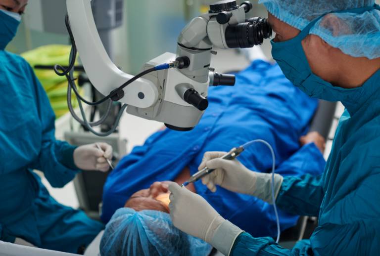
One exciting example of how advancements in engineering are being targeted at a specific clinical problem is the use of robotics in retinal stem cell delivery. Progress in regenerative and cellular therapies has enabled researchers to grow new retinal cells that can be transplanted into the eye. More details about the regenerative process can be found at the London Project to Cure Blindness led by Prof Lyndon Da Cruz and Prof Pete Coffey. This is a hugely significant clinical advancement, however its impact cannot be realised without an effective delivery method.
Currently, delivery is through a hand-held needle which is very technically challenging with limited visual data to support the surgeon. UCL researchers are developing a novel robotic system which will be used during the surgery to steady motion and ensure sub-micrometre precision and manipulation of delicate retinal tissue. The project is a joint venture involving UCL Institute of Ophthalmology, Moorfields Eye Hospital, the Institute of Healthcare Engineering and Wellcome EPSRC Centre for Interventional and Surgical Science (WEISS). It is a truly multidisciplinary collaboration involving scientists, engineers and clinicians.
The engineering lead on this project, Dr Christos Bergeles, emphasised the importance of collaboration, saying “This kind of research cannot be worked on in isolation; its process is reliant on open communication between engineers and clinicians to identify what the real needs and challenges are. Through collaboration both sides are able to anticipate possible constraints to the research and work to solve them. For instance, the pipette that the surgical robot delivers the cells with may be designed primarily by the engineers, but without close input from clinicians and biologists then its diameter may be too small or its pressure flow too high and the cells may be deformed. Multidisciplinary interplay is absolutely integral to the research’s success”.
A different project that also demonstrates the interdisciplinary benefits of applying advancements in engineering to ocular research is a biometry-based concentric tube robot. This robot aims to assist panretinal surgical interventions. Conventional techniques for vitreoretinal surgery are particularly risky because the size and complexity of the human eye requires micro-precise, dexterous surgical manoeuvres. Entrance is made through tiny incisions, limiting the safely reachable workspace within the eye and frequently resulting in scleral stress or lens damage from collision with tools. The robot is comprised of sub-millimetre concentric tubes and aims to overcome these factors. Its ‘snake-like’ design allows for flexible and precise navigation with limited motion at the incision point and reduced risk of lens collision.
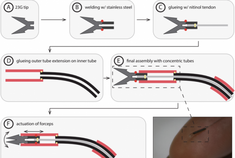
 These are just two examples of the ways in which innovative advancements in robotics technologies are being applied to augment the capabilities of vitreoretinal surgeons and help solve clinical challenges. This a rapidly growing area of ocular research and its strength is built from the kind of close, multidisciplinary collaboration displayed in these projects.
Imaging of the eye
Improvements in imaging the eye and its structures is another area of ocular research that has benefited from advances in engineering technologies. Due to the slow progression and preventable nature of many eye diseases, early diagnosis has proved incredibly effective in increasing the chances of intervention successfully saving patients’ vision. One of the issues however is that by the time loss of sight is identified in conventional eye tests there is often already permanent damage . Adaptive optics scanning light ophthalmoscopy (AOSLO) is an imaging modality that corrects for aberrations of the eye to produce high-resolution images of the eye’s mosaic of cone photoreceptors. The precision of these images allows for the number and arrangement of photoreceptors to be measured, which is a significant biomarker for the early detection of retinal diseases.
Crucially, images produced by AOSLO are typically graded manually by experienced observers. This is very time-consuming and can take several hours per patient, limiting the practicality of this imaging technology. A team of researchers from UCL Institute of Ophthalmology and Moorfields Eye Hospital are developing a machine learning algorithm that can automatically detect, count and classify the photoreceptors instead. The algorithm leverages the appearance of split-detection images to create a cone model that is used for classification. This allows for unsupervised automated detection and has the potential to enable the early detection of retinal pathologies in a far greater number of patients.
Novel imaging techniques are being used within ophthalmology in many different ways. One research project led by Prof Sobha Sivaprasad at Moorfields Eye Hospital aims to tackle blindness caused by diabetic retinopathy, a complication of diabetes in which the eye’s blood vessels are damaged by high blood pressure and high blood sugar levels. This research will evaluate cost-effective imaging measures that would help screen for diabetic retinopathy and allow for early detection. Another area of research addresses the difficulties of keeping cameras focused when imaging eyes due to movement. Research by Dr Pearse Keane is developing an instrument that will use multi-focus plenoptic cameras to reconstruct focused 3D images and overcome the limitations caused by motion. This will greatly enhance imaging capabilities inside the eye and has a potentially wide application for a range of diagnostic and monitoring uses.
What kind of support and infrastructure exists for ocular research at UCL?


The ocular research being developed at UCL occurs within an incredibly interdisciplinary framework. The UCL Institute of Ophthalmology works in close partnership with Moorfields Eye Hospital and NIHR Moorfields Biomedical Research Centre. Together they conduct cutting-edge research with a real patient impact. In addition, the Institute of Healthcare Engineering’s flagship programme Eyes for Life creates a common network of support and infrastructure for researchers. The flagship’s Sandpit events are a chance to bring ophthalmologists and engineers together to stimulate cross-pollination of ideas and project proposals. Read more about our latest Sandpit here.
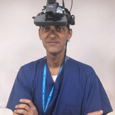
The flagship’s engineering lead Dr Christos Bergeles (top photo) said, “The human eye is an incredibly complex and sensitive organ, built up of tiny micro-structures. As an engineer, I am attracted to these complexities and find it incredibly fascinating (and at times frustrating!) to work with. But ultimately my work is about making an impact on patients. Our research is always aimed at delivering patient-focused results and finding solutions that will benefit the realities of clinical need”.The focus on developing patient-focused results is evidenced through Dr Bergeles work with patient support groups and charities which allow him to disseminate early stage research findings and gather crucial feedback. The close ties between engineers and clinicians, such as with the flagship’s clinical lead Prof Lyndon Da Cruz (bottom photo), means that research is kept tapped into patient need and clinical reality as closely as possible.
To find out more about the research discussed and hear about some of the other advancements being made in ophthalmology, please come join us at New Scientist Live.
New Scientist Live exhibition, London ExCel, 28 September 2017-01 October 201, Stand 1229.