 Close
Close

Image reconstruction tool allows researchers to unlock historic MS data
14 February 2018
A new image reconstruction tool has been developed to support a landmark longitudinal study of 132 multiple sclerosis (MS) patients.
The study has been tracking a group of patients since the 1980’s and the MRI scans from this time, held at the National Hospital for Neurology and Neurosurgery (NHNN), are some of the earliest on record. As the files are held on film and pre-date digital archives, modern image analysis techniques which help clinicians to spot changes in brain structure and volume could not be applied.
The Translational Imaging Group (Institute of Healthcare Engineering) developed a solution to help the team access long-term pathological and morphological evolution from the invaluable early data to perform a fully comprehensive study.
First, the team had to convert each of the 300 sets of historic films into a digitized format, then create reconstructed versions of the scans showing a consistent, volumetric representation. To do so they created a novel algorithm which also helped combat some of the detrimental effects present in archaic films, by sharpening blurred anatomical features and enhancing image contrast to produce good quality data.
Following volumetric reconstruction the research teams now plan to apply modern image analysis methods to spot potential disease and symptom development patterns.
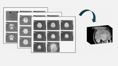
Image Caption: Volumetric image reconstruction from printed MR films. Printed films allow a visual assessment of the brain anatomy on a slice-by-slice basis only. A volumetric reconstruction step recreates a consistent 3D, digital image of the brain from historical scans and unlocks the full potential of modern image analysis tools to understand disease progression in MS.
The study in question aims to enhance our understanding of disease progression in MS and allow researchers to learn more about the relationship between important biomarkers, such as brain and lesion volume, and clinical outcomes.
The study is also unique in being the longest longitudinal study of MS patients with radiological data ever conducted. The earliest patient scans predate the availability of treatment for MS, meaning that researchers can analyse the disease progression of an untreated cohort in a way that is no longer possible. The study therefore offers a unique insight into the relationship between early and intermediate radiological features, and long-term outcomes.
The reconstruction method could allow for retrospective analysis of historical imaging data to take place, further improving our understanding on a range of other disease models. The process of reconstruction has been detailed in full in a recently published paper, Volumetric reconstruction from printed films: Enabling 30 year longitudinal analysis in MR neuroimaging and the framework used is open source and available on GitHub.
This work is a collaboration between the Queen Square Multiple Sclerosis Centre (Institute of Neurology), the National Hospital for Neurology and Neurosurgery and the Institute of Healthcare Engineering (Translational Imaging Group).