 Close
Close

The sensory epithelia of the inner ear are composed of a regular mosaic of hair cells, interspaced by supporting cells. These two cell types derive from a pool of sensory-competent (or prosensory) cells, which are produced at early stages of ear development. We are studying the genes and signals that control the formation of these cells, using a combination of approaches and animal models.
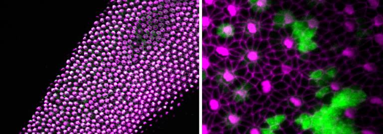
On the left, surface view of a normal basilar papilla - the auditory epithelium of birds. The hair cells have an actin-rich bundle of stereocilia at their surface (in magenta). On the right, a high mag view of a sample in which the Notch ligand Delta1 is overexpressed along with a green-fluorescent protein reporter in some cells...
Most of our research is somehow connected to the roles of the Notch signalling pathway, one of the major cell-to-cell communication systems. Notch has two distinct roles in the developing inner ear. At early stages, it promotes ‘prosensory specification’. In other words, it instructs some cells of the embryonic inner ear (the otic vesicle) to form the parent (precursor) cells for the sensory organs. At later stages, Notch assigns to these cells a hair cell or supporting cell identity by lateral inhibition. This process is very important to ensure that a harmonious balance of hair cells and supporting cells is produced. Our research aims to understand how Notch exerts these different functions in the inner ear: what are the specific genes targeted by Notch at different stages of ear development? How do they contribute to sensory cell or non-sensory cell differentiation.
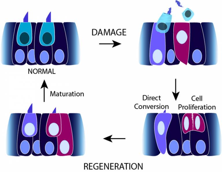
We are also working on the mechanisms of hair cell regeneration – in fact, all non-mammalian species have the remarkable capacity to produce replacement hair cells throughout life. Identifying the factors that allow this regeneration to occur in birds, fish, or amphibians could help designing better cures for hearing loss in humans.
Collaborators: Jonathan Gale, Jonathan Ashmore, Andy Forge (Ear Institute), Vincent Plagnol (UCL Genetics Institute), Jennifer Stone (U. Seattle, USA), Domingos Henrique (U. Lisboa, Portugal), Mike Lovett (Imperial College London), Mark Warchol (Washington U., USA)
Publications:
- ADAM10 and gamma-secretase regulate sensory regeneration in the avian vestibular organs. Developmental Biology 2017
- Costa, A., Sanchez-Guardado, L., Juniat, S., Gale, J.E., Daudet, N., Henrique, D. (2015). Generation of sensory hair cells by genetic programming with a combination of transcription factors. Development (Cambridge), 142 (11), 1948-1959. doi:10.1242/dev.119149
- Eddison, M; Weber, SJ; Ariza-McNaughton, L; Lewis, J; Daudet, N; (2015) Numb is not a critical regulator of Notch-mediated cell fate decisions in the developing chick inner ear. Front Cell Neurosci , 9 , Article 74. 10.3389/fncel.2015.00074.
- Chrysostomou, E., Gale, J.E., Daudet, N. (2012). Delta-like 1 and lateral inhibition during hair cell formation in the chicken inner ear: evidence against cis-inhibition.. Development (Cambridge), 139 (20), 3764-3774. doi:10.1242/dev.074476
- Freeman, S.D., Daudet, N. (2012). Artificial induction of Sox21 regulates sensory cell formation in the embryonic chicken inner ear.. PLOS ONE, 7 (10), doi:10.1371/journal.pone.0046387
- Bird, J.E., Daudet, N., Warchol, M.E., Gale, J.E. (2010). Supporting cells eliminate dying sensory hair cells to maintain epithelial integrity in the avian inner ear.. J NEUROSCI, 30 (37), 12545-12556. doi:10.1523/JNEUROSCI.3042-10.2010
- Daudet, N., Gibson, R., Shang, J., Bernard, A., Lewis, J., Stone, J. (2009). Notch regulation of progenitor cell behaviour in quiescent and regenerating auditory epithelium of mature birds.. DEV BIOL, 326 (1), 86-100. doi:10.1016/j.ydbio.2008.10.033
- Daudet, N., Ariza-McNaughton, L., Lewis, J. (2007). Notch signalling is needed to maintain, but not to initiate, the formation of prosensory patches in the chick inner ear. DEVELOPMENT, 134 (12), 2369-2378. doi:10.1242/dev.001842