Close
Close

FaceValue is a multidisciplinary research team of clinicians, engineers and computer scientists based at UCL GOS Institute of Child Health & Great Ormond Street Hospital for Children.
Great Ormond Street Hospital for Children is a world class healthcare provider with the responsibility of caring for children with the most complex medical challenges of today and the future. Our patients often present with rare and unique anatomical and clinical conditions which demand the highest level of precision in care that is available.
Our overall research objective is to develop novel techniques and devices to understand and improve clinical outcomes and safety of craniofacial surgery for children born with conditions affecting their head and/or face.
We lead five major research themes:
1. 3D Morphometrics
2. Cranial Biomechanics
3. Surgical Devices
4. 3D Printing
5. Clinical Outcomes
You can explore our work by expanding the sections below on this webpage
- 3D Morphometrics
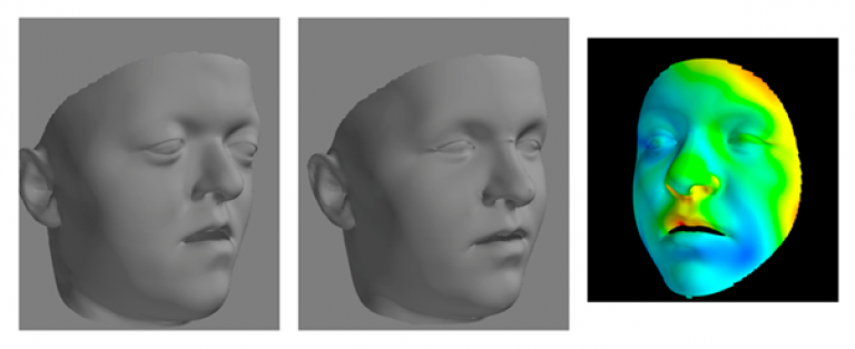
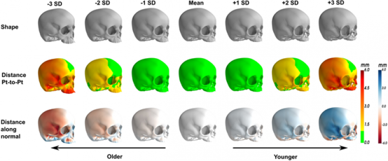
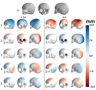
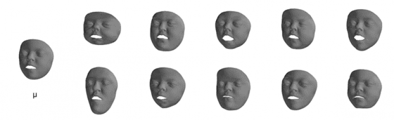
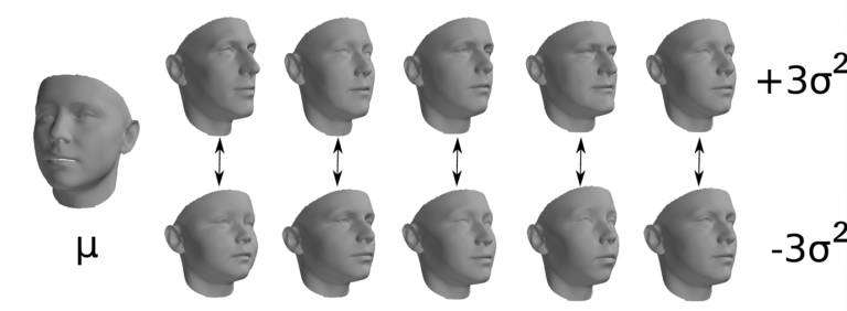
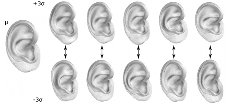
Morphometrics is the quantitative study of form. Traditionally, craniofacial morphometrics has involved measuring distances, angles and ratios between well-defined anatomical landmarks on the face and craniofacial skeleton. It has been an important tool in both medicine and anthropology, as has helped us to understand shape and variations in facial form, but provides little information about 3D shape.
With the advent of sophisticated 3D imaging techniques, computer vision and machine learning, the study of geometric morphometrics (GM), which captures 3D shape allows the construction of detailed statistical models of craniofacial form.
The morphometric group within Face Value is a collaboration between clinicians, engineers and computer vision scientists established to investigate normal and abnormal craniofacial form. Our interests include using GM for
• Machine learning based diagnosis of craniofacial conditions
• Surgical planning and design of new operations to correct craniofacial deformity
• Assessment of surgical outcomes
Machine learning requires large amounts of data. In 2012, our MEIN3D project with the London Science museum collected 12,000 3D face images from the general population and allowed us to construct the world’s largest statistical model of human facial form (LSFM).
Since then, we have constructed models of facial form of children and people with various craniofacial conditions, and are currently developing a statistical skull model.
Our machine learning based diagnostic techniques currently outperform human experts for syndromic craniosynostosis diagnosis.
An academic license for LSFM is available at: https://xip.uclb.com/i/healthcare_tools/LSFM.html
- Cranial Biomechanics
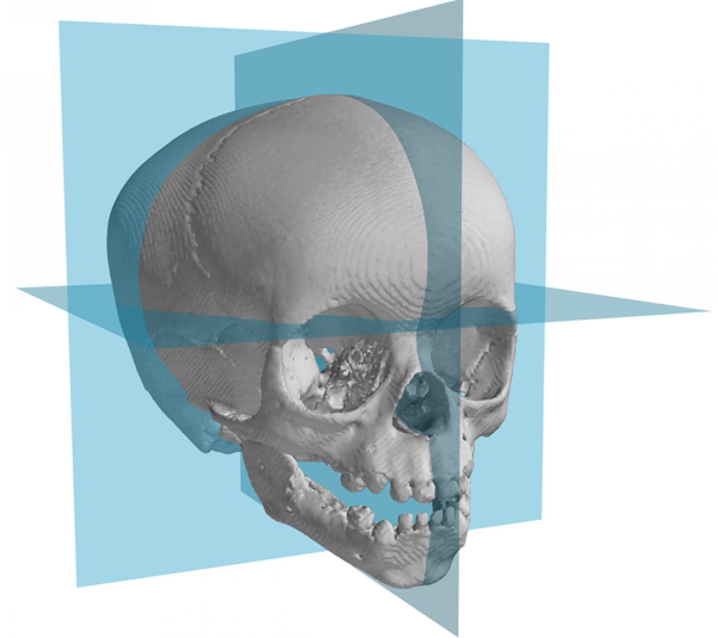
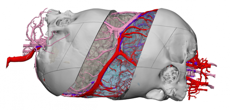
Our research focuses on understanding the mechanical and morphological properties of the foetal and paediatric human cranium using high resolution imaging and experimental techniques. We study the bone physical quality at different stages of skull development and the microstructural parameters that are best suited as biomarkers for prediction, identification and monitoring of congenital conditions such as craniosynostosis. This would support more effective surgical planning and treatment outcomes.
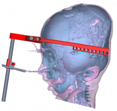
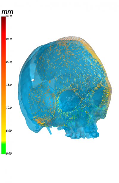
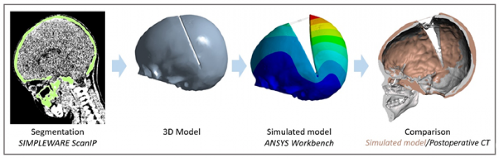
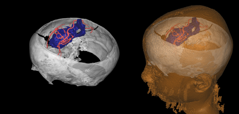
A key research area is in-silico medicine with the goal to develop realistic computational models of craniofacial surgeries to aid clinicians in surgical planning of complex operations.
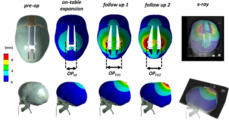
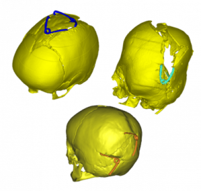
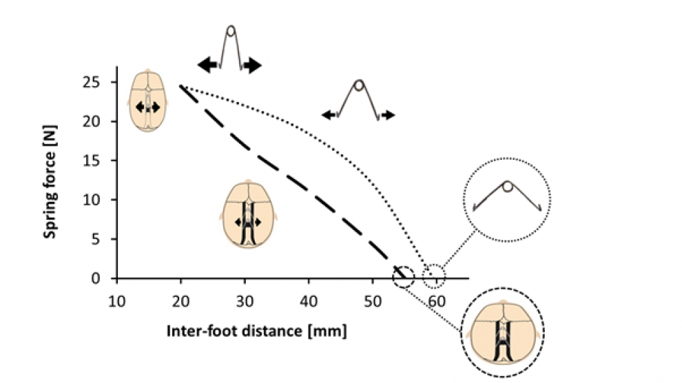
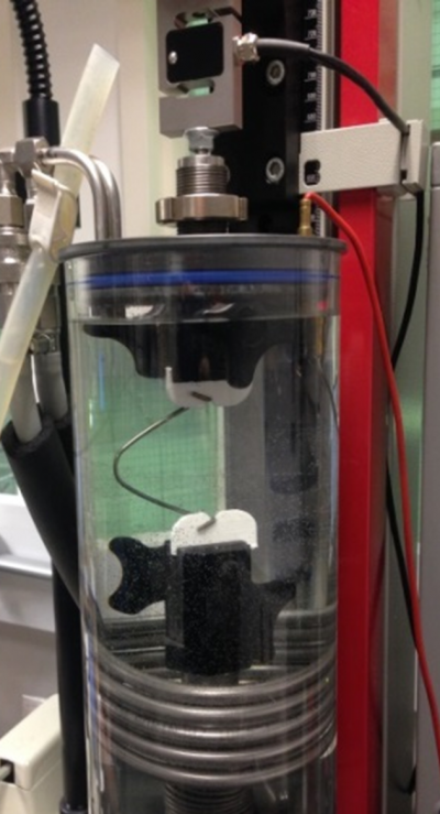
Experimental research tightly coupled with computational studies has enabled us to predict the response of the paediatric calvarium to cranial distractors. We have created and optimised a computational (finite element) model for the prediction of spring outcomes in scaphocephaly correction and spring-assisted posterior vault expansion (SAPVE) outcomes, using routine pre-operative computed tomography (CT) scans.
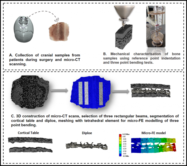
We have investigated the mechanical properties of cortical table and diploe in the parietal bone of patients affected by sagittal craniosynostosis and compared them with microstructural features. Understanding these properties would not only provide insight into bone response to surgical procedures, but also improve the accuracy of our computational models
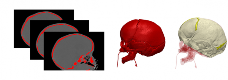
To further understand craniosynostosis bone development, we use a mouse model of Crouzon syndrome that phenotypically mimics human craniosysnostosis.
This model aims at (i) investigating microstructural changes in the cranial bone as a result of FGFR2 mutation during embryonic and postnatal development and (ii) evaluating bone microarchitecture at different stages of skull development.
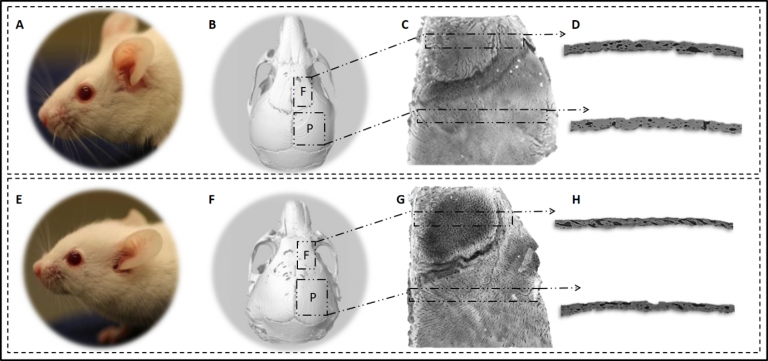
Analysis of the cranial microstructure in the Crouzon mouse model. (A, E) Craniofacial appearance of wild type and heterozygous (Crouzon) mice. (B, F) Visualisation of the regions of interest: frontal bone and parietal bone. (C, G) Micro-CT images of the right side of calvarium. (D, H) High-resolution synchrotron scans of frontal and parietal bone for lacunar analysis. f, frontal bone; p, parietal bone
- Surgical Devices
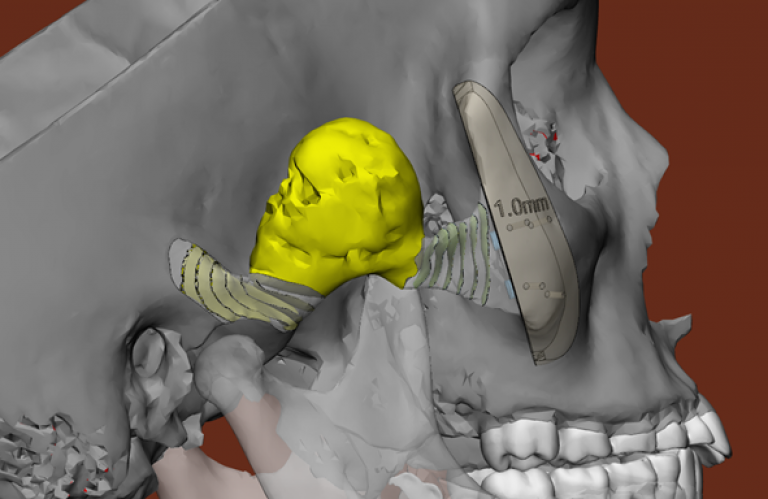
The best option for the patient is not to require any surgery, but until medical technology gets there, our passion at FaceValue is to design surgical devices that make the surgery smaller, safer and more efficient.
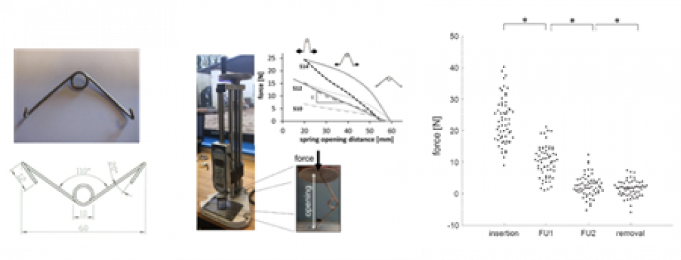
Our knowledge of morphometrics, modelling and materials puts us in the unique position to design such bespoke distractor devices.
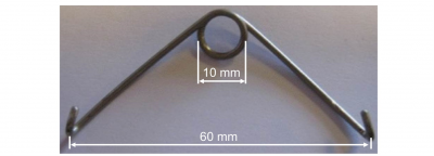
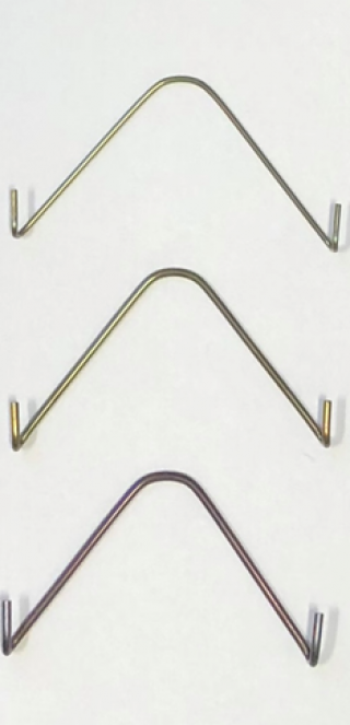
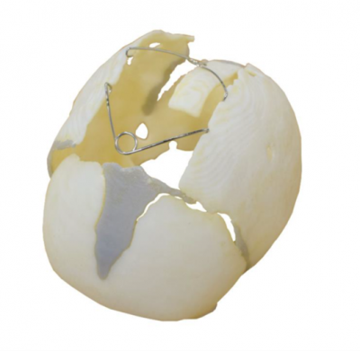
We have pioneered the development of the CranioXpandTM, a surgical spring distraction system in partnership with KLS Martin, Germany. These spring devices alter and treat craniofacial pathologies through minimally invasive procedures. To cite one example, the surgical time for the treatment of scaphocephaly using these devices compared to the traditional operation, is reduced from 150 minutes to <60 minutes; transfusion rates are reduced from nearly 100% to 7% and hospital stay from median 4 nights to 1 night.
We use finite element modelling techniques to plan the surgery beforehand to optimise outcomes in each patient.
Our team is currently working on the second generation of spring distraction devices with shape memory metals and biodegradable materials, to further improve the displacement accuracy and reduce the surgical burden on the patient.
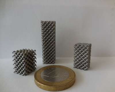
We have designed a mandibular distractor system and shape memory metal meshes using the above technologies.
SS Spring
NITI Spring
NITI Additive Manufacturing
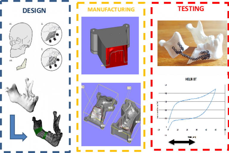
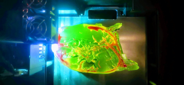
- 3D Printing
Advanced visualisation and rapid prototyping service
Our patients often present with rare and unique anatomical and clinical challenges which demand the highest level of precision in care that is available. Our 3D printing facility serves as a platform which facilitates the understanding of complex 3D problems and group working across multiple disciplines to stimulate innovation and support a range of research themes.
Improved understanding of complex anatomies
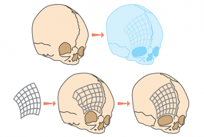
Using 3D printing, we can achieve a greater understanding of rare and complex anatomies which would otherwise be difficult to visualise using conventional 2D slice imaging data. We work closely with radiology to produce accurate & patient specific 3D Printed anatomical models - with the aim to create a better appreciation of the unique organic geometries in question as well as a sense of scale and depth perception.
Enhanced surgical planning
A greater understanding of complex anatomy leads to enhanced surgical planning. We use 3D Printing and virtual surgical planning to improve the quality of care in craniofacial surgery by the reduction of theatre time & costs, increased patient safety, reduced blood transfusions, more accurate resection margins, reduced length of stay and fewer readmissions. The segmented data and 3D models can be integrated with VR/AR solutions which add another degree of interaction with the data.
Improved patient understanding & consent
3D models are being used to help improve communication and understanding of complicated conditions, anatomies and treatments. The physical 3D printed model facilitates discussions between clinicians, patients and parents in a way which is recognisable & easily understood and greatly improves the quality of the consent process
Rapid prototyping
The 3D service provides rapid prototyping capability at the point of care for all clinical specialities. The in-house service improves communication, develops expertise and provides quicker iterations and reduces barriers to the development of novel projects and better informs the final design.
- Clinical Outcomes
Our aim is to ensure that the management of children with craniofacial disorders rests on a firm scientific basis. Treatments are complex and this poses problems for the objective evaluation of results – both functional and appearance change – that the FaceValue Craniofacial Research Unit with its expertise in experimental and computational methodology is uniquely positioned to address. The unit’s extensive clinical experience allows for detailed clinical outcome analysis, aided by advanced measurements tools, following interventions for both single suture synostosis (helmets versus springs, avoiding late deformity, for example) and complex/syndromic forms (benefits versus risk from fronto-facial distraction surgery).