Close
Close

Our group is interested in understanding how embryonic cells develop into different lineages and go on to form a heart during morphogenesis. Understanding cardiac morphogenesis will allow identifying the mechanisms affected in congenital heart diseases -impacting around 1 live birth in every 100- and inform design principles to program tissue patterns and shape for tissue engineering.
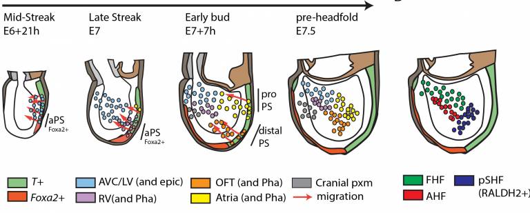
Our heart is composed of four chambers, two ventricles and two atria arranged in parallel and driving blood from the body to the lungs and through the rest of the body. We recently established a fate map demonstrating that each group of cells forming the heart have distinct spatial and temporal origins in the primitive streak (the early embryonic structure that gives rise to mesoderm and endoderm tissues during gastrulation) (PMID: 33999917). The ventricular progenitors are the first to leave the streak, followed by progenitors contributing to the atrium and outflow tract. These observations suggest the cardiac progenitors are prespecified in the primitive streak, prefiguring their allocation to distinct anatomical structures of the heart.
In our latest work, we established new live-imaging methods using light-sheet and multiphoton technologies to enable simultaneous recording of cell migration and intracellular signal transduction pathways during gastrulation in the mouse (see https://www.ivanovitch-lab.com/movies). The analysis is complemented by single-cell RNA sequencing and bar-coding, allowing us to distinguish differentiation trajectories between wildtype and mutant progenitors. We are also investigating if, depending on timing and sites of ingression through the primitive streak, cardiac progenitors adopt distinct migratory routes that expose them to signalling at distinct developmental time points, thus influencing their cardiac fate and morphogenesis.
- Lab Members
Hannah Ford (BHF PhD rotation student)
Isabel North (MSc student- UCL)
Marehiko Kondo (Research Assistant -St Georges University)
Moaz Tallat (Crick-UCL visiting PhD student)
Shayma Abukar (BHF PhD student)
- Selected Publications
Original Articles:
1.Shayma Abukar, Peter Embacher, Alessandro Cicarrelli, Sunita Varsani-Brown, Jamie A Dean, James Briscoe, Kenzo Ivanovitch* Live-imaging reveals Coordinated Cell Migration and Cardiac Fate Determination during Mammalian Gastrulation. bioRxiv 2023.12.19.572445; doi.org/10.1101/2023.12.19.572445
2. M. Joaquina Delás, Christos M Kalaitzis, Tamara Fawzi, Madeleine Demuth, Isabel Zhang, Hannah T Stuart, Elena Costantini, Kenzo Ivanovitch, Elly M Tanaka, James Briscoe. Developmental cell fate choice employs two distinct cis regulatory strategies. Developmental Cell 2022.06.06.494792; doi.org/10.1016/j.devcel.2022.11.016
3. Ivanovitch K*, Soro-Barrio P, Chakravarty P, Jones RA, Mousavy Gharavy SN, Stamataki D, Delile J, Smith J.C, Briscoe J. Ventricular, atrial and outflow tract heart progenitors arise from spatially and molecularly distinct regions of the primitive streak. PLOS Biology doi.org/10.1371/journal.pbio.3001200 (2021)
4. Collart C., Ciccarelli A., Ivanovitch K., Rosewell I., Kumar S., Kelly G., Edwards A., Smith J.C. The migratory pathway of the cells that form the endocardium, dorsal aortae, and head vasculature in the mouse embryo. BMC Dev Biol 21, 8 (2021)
5. Metzis V, Steinhauser S, Pakanavicius E, Gouti N, Stamataki D, Ivanovitch K, Watson T, Rayon T, Neda Mousavy Gharavy S, Robin Lovell-Badge, Nicholas M Luscombe and James Briscoe. Nervous System Regionalization Entails Axial Allocation before Neural Differentiation. Cell Oct 2018 Nov 1;175(4):1105-1118.e17.
6. Ivanovitch K, Temino Valbuena Susana, Torres M. Live imaging of heart tube development in mouse reveals alternating phases of cardiac differentiation and morphogenesis. eLife Dec 2017;6:e30668
7. Le Garrec JF, Dominguez JN, Desgrange A, Ivanovitch K, Raphael E, Bangham JA, Torres M, Coen E, Mohun TJ and Meilhac SM. A predictive model of asymmetric morphogenesis from 3D reconstructions of mouse heart looping dynamics eLife Nov 2017;6:e28951
8. Diz-Munoz A, Romanczuk P, Yu W, Bergert M, Ivanovitch K, Salbreux G, Heisenberg CP, Paluch EK. Steering cell migration by alternating blebs and actin-rich protrusions. BMC Biol. 2016 Sep 2;14(1):74.
9. Ivanovitch K, Cavodeassi F, Wilson SW. Precocious acquisition of neuroepithelial character in the eye field underlies the early onset of eye morphogenesis. Developmental Cell 2013 Nov, (27), 293-305
10. Cavodeassi F, Ivanovitch K, Wilson SW. Eph/Ephrin signalling maintains eye field segregation from adjacent neural territories during forebrain morphogenesis. Development 2013 Oct 140, 4193-4202
11. Picone R, Ren X, Ivanovitch K, Clarke JDW, McKendry RA, Baum B. A Polarised Population of Dynamic Microtubules Mediates Homeostatic Length Control in Animal Cells. PLoS Biol. 2010 Nov 16;8(11)
12. Tamori Y, Bialucha CU, Tian AG, Kajita M, Huang YC, Norman M, Harrison N, Poulton J, Ivanovitch K, Disch L, Liu T, Deng WM, Fujita Y. Involvement of Lgl and Mahjong/VprBP in cell competition. PLoS Biol. 2010 Jul 13;8(7)
13. Fichelson P, Moch C, Ivanovitch K, Martin C, Sidor CM, Lepesant JA, Bellaiche Y, Huynh JR. Live imaging of single stem cells within their niche reveals that a U3snoRNP component segregates asymmetrically and is required for self-renewal in Drosophila. Nat Cell Biol. 2009 Jun;11(6):678-9
Review Article:
1. Ivanovitch, K; Esteban, I.; Torres, M. Growth and Morphogenesis during Early Heart Development in Amniotes. J. Cardiovasc. Dev. Dis. 2017 Nov 22;4(4):20.