Transmission Electron Microscopy
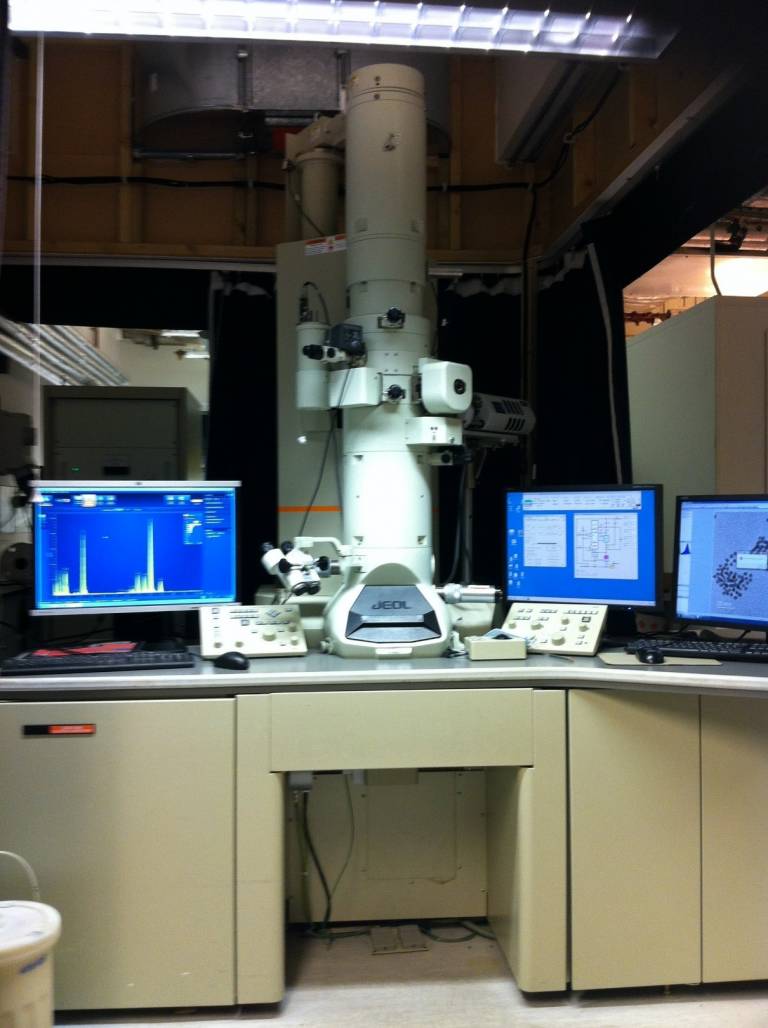
A Jeol 2100 200 kV fitted with a LaB6 filament giving a point resolution of 0.13 nm. It is equipped with bright and dark field STEM detectors and an Oxford Instruments EDS detector. It is housed in LG26, in the basement.
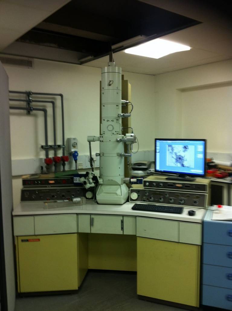
A Jeol CX100 II 100 kV with a tungsten filament. The microscope is nearly 40 years old and can resolve features as small as 2 – 3 nm, although this is very sample dependent. It is housed in room LG27, in the basement.
The 2100 is very heavily used, often booked up 5 – 6 weeks in advance, so it is always quicker to use the 100 CX to screen samples, even if it is not capable of high resolution (e.g. lattice plane) imaging. The 100 CX is much quicker to set up and align, and two sample grids can be inserted together.


For either TEM, it is critically important that the samples are non-magnetic. When the sample is inside the microscope column it is in a powerful magnetic field, so if it is magnetic it will be attracted to the closest magnetic lens, the objective polepiece, and may become stuck there. This magnetic material will then distort the magnetic field of the objective lens, making focusing difficult or impossible. If this happens the TEM will have to be dismantled and cleaned, which is a time consuming and expensive procedure. Also, if the sample is magnetic it won’t be possible to get good images from it because the magnetic field of the sample will distort the trajectory of the electrons, making it impossible to produce a focused image. For these reasons, magnetic materials must NEVER be put into these TEMs. The most common elements that can produce magnetic compounds are iron, cobalt and nickel, so ALL samples containing any of these elements MUST be tested before being inserted into the TEM column. Compounds of more unusual elements, such as gadolinium and neodymium can also be magnetic, so any samples containing these elements MUST also be tested. The testing MUST be done on the bulk sample, not the sample on the grid. There is a neodymium magnet in the SEM room, LG27, and this can be used to test the sample. If any samples containing these elements is put into the TEM column without being tested, the user will be banned from using the TEMs, even if the sample is subsequently found to be non-magnetic.
The pressure inside the TEM column is of the order of 10-6 Pa, so the sample must be dry before being inserted into these TEMs.
Samples must be thinner than about 100 nm, or the electrons will not pass through them. This means films on a substrate cannot be imaged, but most powders can be. If the particle size is greater than 100 nm the powder can be sonicated in e.g. methanol or hexane to form a suspension. One or two drops of this suspension are put onto a grid and left to evaporate. Large particles or clusters will just produce a silhouette image, but it is often possible to get images of details at the edges of such large particles, or simply to find smaller particles on the grid.
To put the samples into the TEM, they must be mounted onto a sample grid. These grids are ca. 3 mm diameter circular mesh of copper or gold, with a layer of carbon to support the sample. It is important to put the sample on the darker, carbon coated, side of the grid. Samples are usually prepared by sonicating the powder in a volatile liquid such as hexane or methanol to produce a suspension. Two or three drops of this suspension are dropped onto the dark side of the grid and left to dry. Thin films cannot be imaged directly, but can be scraped off the substrate and the scaping sonicated as above. Alternatively, the film and substrate can be sonicated together, and anything that comes into suspension can be dropped onto the grid as above. Of course, these methods destroy the integrity of the film. When the sample grids are dry, they are mounted onto a sample rod and inserted into the TEM for imaging. The sample rod for the 2100 will only hold one sample grid, so each sample must be imaged individually. The sample rod for the 100 CX can hold two sample grids.
When the samples have been inserted into the TEM they are left for two or three minutes for the pressure in the column to return to the operating pressure. Then the filament is turned on to generate the electron beam and the beam is aligned down the microscope column. These alignment procedures will be shown and explained during the training session. They are different for the two TEMs, so there are separate training sessions for each microscope. For the 2100, the training to prepare and insert the sample, align the microscope and image the sample will take three to four hours. The training will be done on your sample, and the rest of the session can be spent imaging another of your samples. For the 100 CX, the training will take two to three hours, and will be done on one of your samples. The rest of the session can be spent imaging any other samples you may have.
For training, contact s.firth@ucl.ac.uk.
Images are taken off the pcs and saved onto DVDs. This is because the internet cannot be connected to the pcs controlling these microscopes as it would interfere with the communication between the microscope and the controlling software. Therefore there is no antivirus software on these pcs, so USB memory sticks must NEVER be used. DVDs have to be used, and there are always some available in the lab.
 Close
Close

