Close
Close

I am a neuroscientist who is interested in developing and utilising novel technologies to remotely and precisely manipulate brain cells such as astrocytes with magnets and magnetic materials. In particular, I leverage the high-field magnetic resonance imaging (MRI) systems in the Centre for Advanced Biomedical Imaging (CABI) at University College London (UCL) to achieve imaging and actuation with a single device. My current research areas include astrocyte biology and brain cancer therapy.
Research
Magnetomechanical stimulation (MMS)
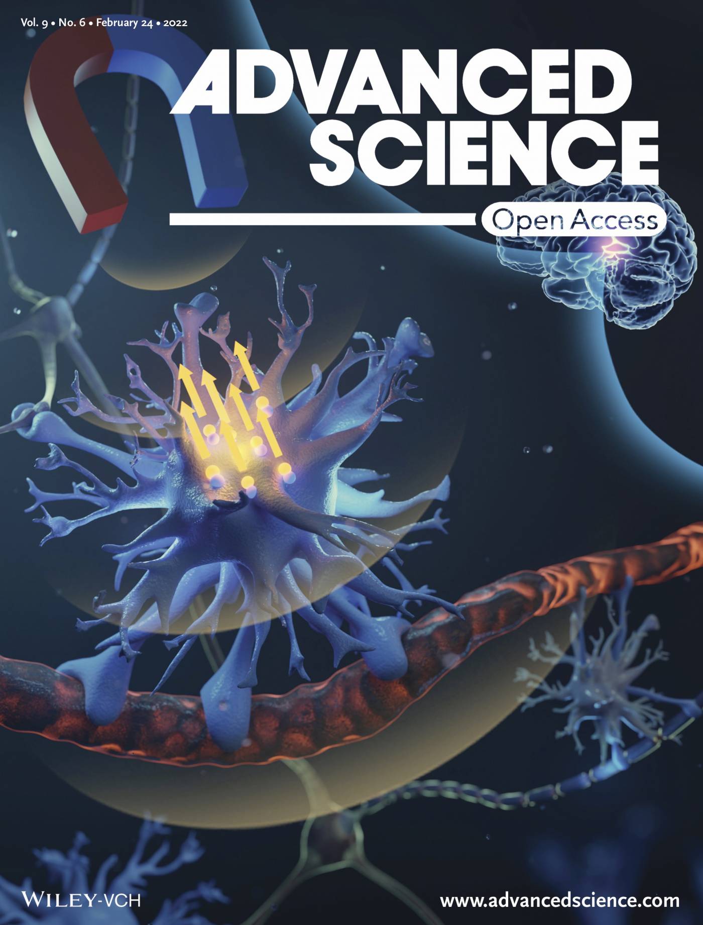
Astrocytes are a major type of glial cells in the central nervous system (CNS). They play crucial and diverse roles in the functioning of the brain, and are implicated in many neurological conditions including neurodegenerative diseases, epilepsy, stroke, and mood disorders. As researchers strive to better understand astrocyte biology and to develop novel, astrocyte-targeting therapies, technologies that enable selective control of astroglial activity in a live brain are increasingly needed. However, existing methods such as optogenetics and chemogenetics require the introduction of foreign proteins into the cells of interest, which adds a layer of complication and hinders their clinical translation.
To overcome this obstacle, I led the development of a novel brain cell stimulation technology called ‘magnetomechanical stimulation (MMS)’ that enables remote and selective control of astrocytes without genetic modification. MMS exploits the intrinsic sensitivity of astrocytes to mechanical stimuli and triggers mechano-gated calcium and adenosine triphosphate (ATP) signalling by applying a magnetic field to antibody-functionalised magnetic particles that are targeted to astrocytes.
First, we determined the minimum mechanical stimulus required to elicit calcium and ATP signalling responses from astrocytes in culture, using a custom-built electromagnet that can apply precise forces to magnetic particles.
We then demonstrated that, after delivering magnetic particles to astrocytes in a live brain, a single MRI system can be used to both image and actuate the particles, resulting in the stimulation of specific astrocytes in the rat brainstem. Imaging allows assessment of particle delivery, and actuation is achieved with the fringe magnetic field of the scanner.
Unlike existing techniques such as optogenetics and chemogenetics, MMS requires neither device implantation nor genetic modification, therefore it does not face these significant obstacles to clinical translation. In addition, the safety of magnetic iron oxide nanoparticles has been extensively studied, both preclinically and clinically, since the 1990's, and they have been shown to have a high degree of biocompatibility in the brain and are well tolerated by astrocytes and neurons. Lastly, the common existence of MRI scanners in hospitals may enable MMS to be implemented with minimal hardware investment and development. All of these features make MMS a promising candidate for development as a neuromodulation therapy.
Minimally INvasive IMage-guided Ablation (MINIMA)
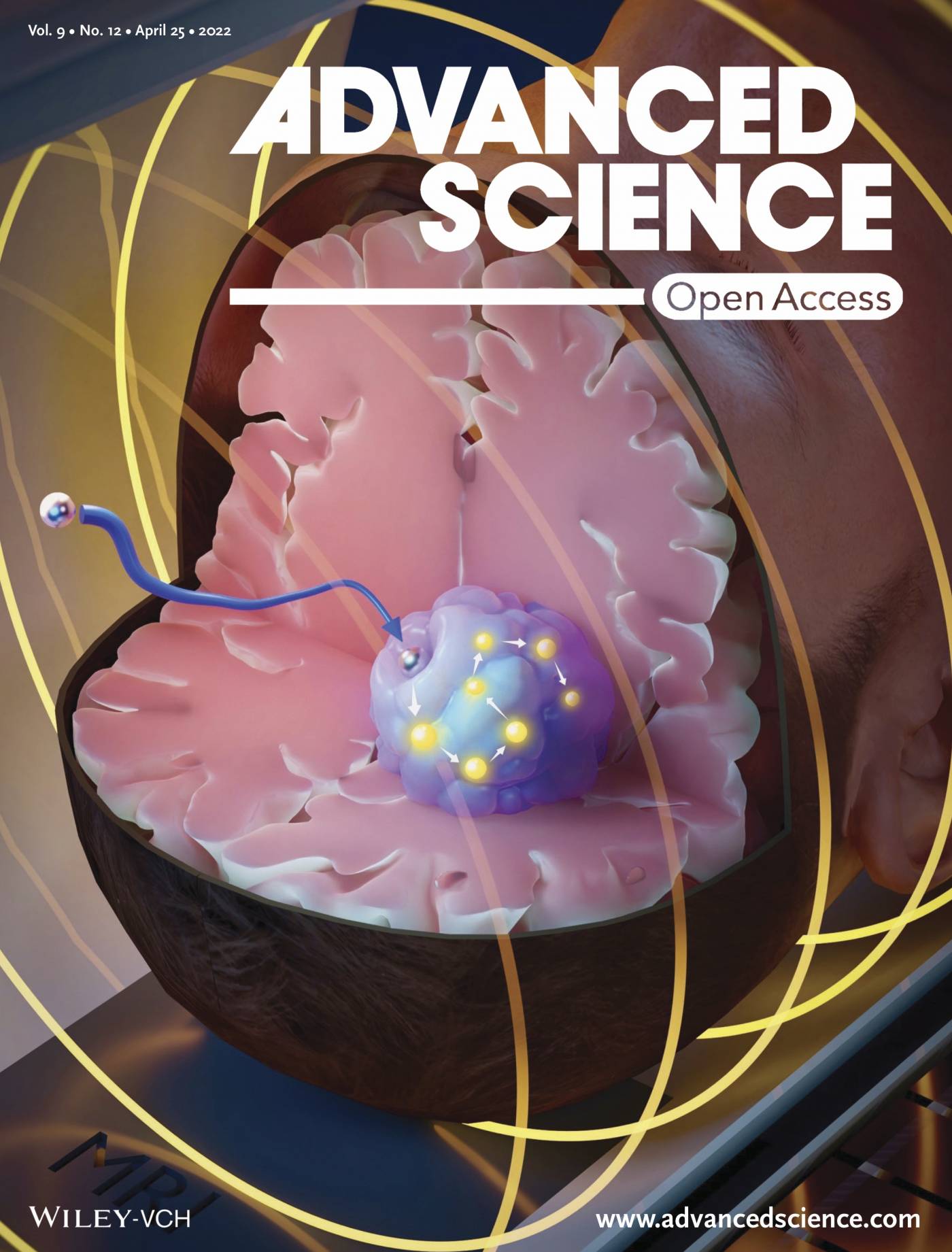
I am also part of the team that developed a novel brain cancer therapy that utilises an MRI system to image, navigate and heat a thermoseed, in order to deliver hyperthermia in a highly controlled manner. The thermoseed is located with a bespoke imaging sequence, steered to the cancerous tissue using the imaging gradients of the scanner, and heated with an MR-compatible radiofrequency coil to cause localised cell death. This process is repeated to ablate multiple tumours or a large tumour, and offers the advantage of fine control over the location and volume of thermablation.
Biography
| 2016 - Present | Centre for Advanced Biomedical Imaging, UCL, UK |
| Research Associate | |
| 2011 - 2018 | Centre for Advanced Biomedical Imaging, UCL, UK |
| PhD in Biomedical Imaging | |
| 2010 - 2011 | UCL, UK |
| MSc in Neuroscience | |
| 2006 - 2009 | University of Cambridge, UK |
| BA in Natural Sciences Tripos | |
Selected publications
Baker, R.R., Payne, C., Yu, Y., Mohseni, M., Connell, J.J., Lin, F., Harrison, I.F., Southern, P., Rudrapatna, U.S., Stuckey, D.J., et al. (2022). Image-Guided Magnetic Thermoseed Navigation and Tumor Ablation Using a Magnetic Resonance Imaging System. Adv. Sci. 2105333.
Yu, Y., Payne, C., Marina, N., Korsak, A., Southern, P., García-Prieto, A., Christie, I.N., Baker, R.R., Fisher, E.M.C., Wells, J.A., et al. (2022). Remote and Selective Control of Astrocytes by Magnetomechanical Stimulation. Adv. Sci. 9, 2104194.
Turovsky, E.A., Braga, A., Yu, Y., Esteras, N., Korsak, A., Theparambil, S.M., Hadjihambi, A., Hosford, P.S., Teschemacher, A.G., Marina, N., et al. (2020). Mechanosensory signaling in astrocytes. J. Neurosci. 40, 9364–9371.
Fratta, P., Sivakumar, P., Humphrey, J., Lo, K., Ricketts, T., Oliveira, H., Brito-Armas, J.M., Kalmar, B., Ule, A., Yu, Y., et al. (2018). Mice with endogenous TDP-43 mutations exhibit gain of splicing function and characteristics of amyotrophic lateral sclerosis. EMBO J. 37, e98684.
Powell, N.M., Modat, M., Cardoso, M.J., Ma, D., Holmes, H.E., Yu, Y., O’Callaghan, J., Cleary, J.O., Sinclair, B., Wiseman, F.K., et al. (2016). Fully-automated μMRI morphometric phenotyping of the Tc1 model of Down syndrome. PLoS One 9, e0162974.
Howard, L.R., Javadi, A.H., Yu, Y., Mill, R.D., Morrison, L.C., Knight, R., Loftus, M.M., Staskute, L., and Spiers, H.J. (2014). The hippocampus and entorhinal cortex encode the path and Euclidean distances to goals during navigation. Curr. Biol. 24, 1331–1340.