 Close
Close

All info from VisualSonics website https://www.visualsonics.com/product/imaging-systems/vevo-2100
Ultrasound with high image quality, increased frame rates, superb contrast, unrivalled resolution and a wide field of view. The system is easy to use, non-invasive and fast, providing extremely high throughput when needed.
Contact: d.stuckey(at)ucl.ac.uk; a.david(at)ucl.ac.uk
Applications
- b-mode for chamber volumes, ejection fraction and LV mass
- m-mode and anatomical m-mode for fractional shortening
- pulse wave Doppler for atrial, aortic and carotid flow
- colour and power Doppler for blood flow quantification & anatomical identification
- needle guidance for closed chest intracardiac injections
- 3D tumour volumes
- Tumour perfusion
Developmental research
- 3D imaging of embryos in situ
- Monitoring brain development
- Measuring cardiac function
- Image guided injections
- 3D visualisation
- Neurobiology
- Blood flow analysis
- Hepatology
- Nephrology
- Ophthalmology
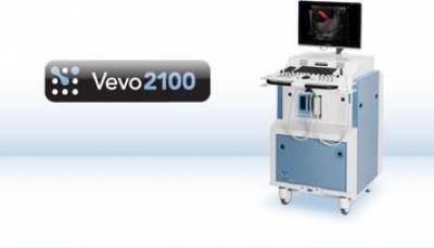
Vevo 2100 Ultrasound system
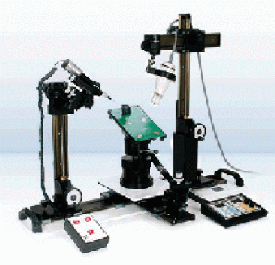
Imaging stage
Imaging modes
|  M-mode imaging of mouse left ventricle |
|  Color Doppler imaging in the mouse spleen |
|  3D B-Mode imaging of orthotopic hepatoma tumor. Dark blue tumor volume. Light blue liver, Orange kidney |
See the collaborations page for information on access to this system