Close
Close

A Single Plane Illumination Microscope (SPIM) is being developed at CABI to record high-resolution, three-dimensional, optical images. Constructed with two lasers, this system will allow illumination at two different wavelengths. The system is being built following the OpenSPIM specification, in collaboration with Pavel Tomancak at the Max Planck Institute of Molecular Cell Biology and Genetics (MPI-CBG), Dresden.
SPIM principle
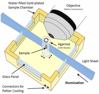
In SPIM, optical sectioning is achieved by illuminating the sample with a sheet of laser light. The specimen is attached to a stage, rotated and translated orthogonally to the light sheet. Image stacks are acquired along multiple directions and then merged into a single three-dimensional data set. The specimen is illuminated from one side and fluorescent light is collected by a detector positioned perpendicularly to the light sheet. Since each plane is illuminated only once, fluorophores outside the focal plane are not exposed to light, and photo damage is therefore dramatically reduced.
SPIM applications
SPIM is a very promising imaging technique that allows the registration of single-cell resolution images of various kinds of samples. Indeed, it can not only be used to examine large cleared specimens, but also to study the development of small insects and animals (drosophila, zebra fish) over several days.
SPIM drosophila embryo
His-YFP drosophila embryo imaged from gastrulation until muscle contractions in late embryogenesis - ventral view. Used with the permission from Pavel Tomancak and Peter Gabriel Pitrone, MPI-CBG
SPIM Management team
| Angela d'Esposito, Main contact |
| Mark Lythgoe, Director of CABI |
| Simon Walker-Samuel, Senior Research Associate |
| Adrien Desjardins, Senior Research Fellow and Lecturer, Medical Physics |
See the collaborations page for information on access to this system