Close
Close

Magnetic Resonance Imaging has revolutionised diagnostic medicine, allowing unprecedented visualisation of internal structure. It has thus rapidly become the clinical gold standard, allowing detailed assessments of pathology and function across the whole body.
Magnetic resonance is a phenomenon that occurs in water protons. Water molecules contain two protons which align with the direction of the magnetic field inside the scanner. Protons are then excited with a radio frequency (RF) pulse which causes their magnetisation to rotate in phase. This magnetic rotation can induce a current in an antenna, which is amplified to form the MRI signal. By timing the applied RF pulses, this signal is made sensitive to the tissue environment in which the water protons reside. This leads to the essential contrast that allows identification of different tissue types. As this approach does not require the use of ionising radiation such as x-rays, repeated in-vivo experiments are possible, which have little effect on the subject.
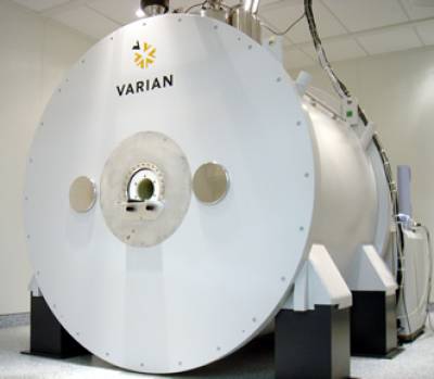
The experimental MRI scanner installed in CABI utilises a much higher magnetic field compared to a typical clinical scanner, allowing us to push the boundaries of anatomical imaging and acquire images with less than 40 micron resolution.
As a team we are committed to developing MRI as a tool to yield information beyond tissue structure. Gated MRI can image the beating heart, as the scanner can be synchronised to an ECG trace. Diffusion weighted imaging can yield structural information on the cellular level. Arterial spin labelling can quantify cerebral perfusion.
We also have extensive experience in the field of molecular imaging using MRI. By using antibodies attached to iron oxide particles we can sensitise the respective receptor distribution to be detectable on an MRI image. For example we can track the progression of tumor pathology. A similar approch can be used to investigate cellular dynamics.
Our aim is to use all of these techniques to non-invasively investigate the complex relationship between structure, function, disease processes and therapy.
We are always interested in expanding our imaging to new applications and finding collaborations that provide opportunities to employ and develop our techniques.
See the collaborations page for information on access to this system