Close
Close

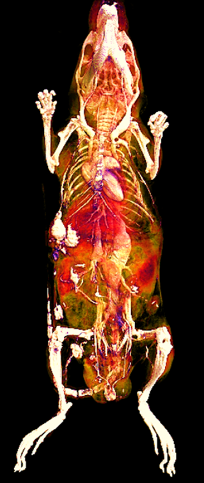
X-ray computerized tomography (X-ray CT) is widely available and is the most frequently used imaging technique in the clinic. CT images are generated by combining series of two dimensional X-ray images taken from multiple projections around a single axis using tomographic reconstruction. CT imaging can provide more informative cross-sectional images and high resolution three dimensional representation of an object. Therefore CT is routinely combined with other tomographic nuclear techniques such as positron emission tomography (PET) or single-photon emission computed tomography (SPECT) to provide 3D anatomical localisation.
At CABI we have developed our CT subsystems to be stand-alone systems with novel applications. CT imaging is a widely used imaging technique to study lung and pulmonary diseases due to natural contrast between air filled lungs and soft tissue. The ability of creating contrast in CT is based on the fact that different tissues provide different degree of X-ray attenuation. For example, bone can provide positive contrast in CT image due to its high X-ray attenuation. Therefore CT imaging is very useful to study bone morphology and density. Other ways of creating contrast in CT is via contrast agents. At CABI we have been investigating gold nanoparticles as a CT contrast agent. Recent works shows that gold can provide high resolution images of the vasculature and vascular remodelling present in disease. We are now applying this to other applications such as cell tracking.
See the collaborations page for information on access to this system