Close
Close

The Microscopy and Imaging Translational Technology Platform provides access to cutting-edge imaging instrumentation to researchers at the UCL Cancer Institute and within the CRUK UCL Centre.
The Translational Technology Platform (TTP) houses a number of microscopy instruments, including instruments capable of long-term time-lapse, laser microdissection and super-resolution, to name a few. The diverse range of instruments allows for flexibility in experiment design and implementation.
The TTP is supported by a dedicated Translational Technology Platform Manager, who can contribute to projects by assisting with both the experimental strategy and design of experiments, as well as implementing techniques that require expert knowledge and skills to develop innovative and novel methodologies.
New users and booking
All new users must be trained on the instruments. You will be required to undertake a 1 hour induction session which will be charged at the hourly rate. The induction will demonstrate all capabilities of the instrument and give the user an opportunity to ask specific application questions. Hands on help and advice can be provided to all users,as well as assistance in experimental strategy and design. Access to relevant booking information will be provided once training is complete.
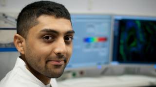
To organise a suitable time for an induction, please contact Jiten Manji (ci.microscopyandimaging@ucl.ac.uk).
Following appropriate training, you will be given access to the relevant booking calendars on the Clustermarket Bookkit system.
Instruments and specifications
- 3i Spinning Disk + TIRF
Spinning Disk Confocal microscope with TIRF and Laser Ablation, Slidebook
This instrument has 4 laser lines, 2 x sCMOS sensors (for Spinning Disk) and 1 x sCMOS sensor (for TIRF) and is capable of imaging up to 40fps. It is fully equipped for time-lapse imaging.
Objectives
10x, 20x, 40xOil, 63xOil, 100xOil (TIRF capable) all Plan-Apochromat
Laser lines
405nm, 488nm, 561nm, 640nm
355nm (ablation)
Image Acquisition
Spinning Disk: 2 x sCMOS Prime95B
TIRF: 1 x sCMOS Prime95B
Peripherals
- Piezo stage
- Definite focus 2
- Environment chamber – CO2 control
- Temperature control
3i Marianas Product Information - https://www.intelligent-imaging.com/marianas
A charging structure will be put in place for use of this machine. Please contact Jiten Manji for further information.
- Zeiss LSM 880 with AiryScan
Confocal microscope, Zen Black
This instrument has 7 laser lines, 2xPMT detectors and 1x32 channel GaAsP detector and is capable of imaging to a resolution of 130nm. It is fully equipped for time-lapse imaging.
Objectives
5x, 10x, 20x, 40xOil, 63xOil all Plan-Apochromat
Laser lines
405nm ,458nm, 488nm, 514nm, 561nm, 594nm and 633nm
Image Acquisition
2xPMT
1x 32 channel spectral GaAsP
1x 32 element GaAsP
Peripherals
- Piezo stage
- Definite focus
Zeiss LSM 880 product information
- Environment chamber
- Temperature control
- Heated stage inserts
A charging structure will be put in place for use of this machine. Please contact Jiten Manji for further information.
- Zeiss LSM900
Confocal microscope, Zen Black
This instrument has 4 laser lines, 2xGaAsP detectors and is dual colour imaging enabled. It is equipped with a scanning stage
Objectives
10x, 20x, 40xOil and 63xOil all Plan-Apochromat
Laser lines
405nm, 488nm, 561nm, 647nm
Image Acquisition
2xGaAsP detectors
Peripherals
Scanning Stage
A charging structure will be put in place for use of this machine. Please contact Jiten Manji for further information.
- Nikon Biostation CT
Long-term time lapse, NIS-Elements
The Biostation has a built in incubator allowing researchers to monitor their cells over long periods of time. It is also able to image up to three fluorescent channels.
Objectives
2x, 5x, 10x, 20x and 40x all phase capable
Fluorescence cubes
CFP, GFP, YFP, DsRed and Cy5
Image Acquisition
CMOS monochrome camera
Peripherals
Incubation temperature control, CO2 control, Humidity control and O2 concentration control Adapters to allow imaging of well plates (any format), dishes and flasks. Nikon Biostation CT product information- Perkin Elmer Vectra 300
Automated imaging cytometer, Phenochart & InForm
This instrument allows for automated slide scanning and spectral unmixing of up to 6 fluorescent parameters. It can scan up to 200 slides in one session.
Objectives
5x, 10x, 20x and 40x all long working distance and phase capable
Fluorescence cubes
DAPI, GFP, TxRed and Cy5
Image Acquisition
Monochrome CCD and Colour CCD cameras
Peripherals
XY scanning stage Automated functionality
- IncuCyte Zoom
Medium-term time lapse, IncuCyte Vesselview
The Zoom is a compact microscope purpose built to image from inside a temperature controlled incubator. It allows for medium-term time lapse imaging with up to two fluorescence parameters.
Objectives
4x, 10x, 20x
Fluorescence
Green: Ex 440-480nm, Em 504-544nm
Red: Ex 565-605nm, Em 625-705nmImage Acquisition
Inverted microscope kept at 37oC on a 2-hour imaging schedule.
Peripherals
96-well scratch maker
- IncuCyte FLR
Medium-term time lapse, IncuCyte Vesselview
The FLR is a compact system designed to image from within a temperature controlled incubator with up to one fluorescent parameter.
Objectives
4x, 10x
Fluorescence cubes
GFP
Image Acquisition
Colour CCD
- Zeiss Live Cell Imager
Short-term time lapse, Axiovision
This instrument has temperature, CO2 and O2 control. It provides high resolution for short-term time lapse imaging experiments.
Objectives
10x, Long Distance 20x, 20x, Long Distance 40x, 40x and 100x Oil
Fluorescence cubes
DAPI/GFP/CFP CFP/RFP
Peripherals
Colibri LED fluorescence illumination XY Scanning stage Environmental Chamber Controlled atmosphere – Temp, CO2 Heated plate inserts
Image Acquisition
Axiocam MRm – Fluorescence
- Zeiss Z1 Inverted with Apotome
Widefield inverted epifluorescent microscope, Axiovision
This instrument is a motorised inverted epifluorescent microscope, capable of imaging up to five fluorescent parameters. The Apotome attachments allows for Z sectioning for thicker specimens.
Objectives
5x, 10x, Long Distance 20x, 20x, 40x and 100x Oil
Fluorescence cubes
DAPI, GFP, Cy5, PI, TxRed
Peripherals
Zeiss Apotome – Structured illumination
Image Acquisition
Axiocam MRm – Fluorescence
- Zeiss A1
Epifluorescent microscope, Axiovision
The A1 is an upright microscope with two cameras available: a monochrome camera for fluorescence and a colour camera for chromogenic staining.
Objectives
5x, 10x, 20x, 40x, 63x Oil, 100x Oil
Fluorescence cubes
DAPI, GFP, Rhod, tdTom
Image Acquisition
Axiocam MRm – Fluorescence Axiocam HRc – Colour
- Zeiss PALM Microbeam
Laser microdissection and capture, PALM Robo
Based on a Zeiss Z1 system, this instrument utilises a 355nm laser to dissect tissue and capture tissue contact free, reducing potential contamination.
Objectives
5x, 10x, 20x, 40x, 63x and 100x Oil
Fluorescence cubes
DAPI, GFP, DsRed
Image Acquisition
Axiocam MRm – Fluorescence Axiocam iCC fast - Colour
Peripherals
XY scanning stage 3 Slide holder, petri dish holder, single and double capture tube holders
- Evos FL Auto
Epifluorescent microscope, EvosFL Auto
This epifluorescent microscope can image up to three fluorescent parameters. The automated stage allows for scanning tissue sections and stitching images.
Objectives
5x, 10x, 20x and 40x all long working distance and phase capable
Fluorescence cubes:
DAPI, GFP, TxRed and Cy5
Image Acquisition
Monochrome CCD and Colour CCD cameras
Peripherals
- XY scanning stage
- Automated functionality
- Leica M205FA
Stereo microscope, Leica LAS
The M205FA is a versatile instrument which allows researchers ample working room between the objective and the specimen. It can image up to 3 fluorescent parameters.
Objectives
Apochromatic variable – 7.8x to 160x
Fluorescence cubes
DAPI, GFP, RFP
Image Acquisition
Leica DFC310 – Colour
- Zeiss Z1 Upright
Epifluorescent microscope, Zen Blue
This instrument is a motorised upright epifluorescent microscope, capable of imaging up to five fluorescent parameters. Zen blue is used as the acquisition software.
Objectives
2.5x, 5x, 10x, 20x, 40x
Fluorescence cubes
DAPI, GFP, Cy5, PI, TxRed
Image Acquisition
Axiocam MRm – Fluorescence
- Image Analysis
Available packages:
- Imaris Full Licence
- Nikon NIS Elements AR
- Zeiss Zen Black deconvolution
Services
- Basic Training for Zeiss LSM 880 with Airyscan
Training will last 1 hour and will go through the basic functions of the instrument and Z-stacking. Users will be charged 1 hour at the instruments hourly rate. Please contact Jiten to arrange this.
- Advanced Training for Zeiss LSM 880 with Airyscan
This will last 1 hour and will go through user specific applications for the instrument. Techniques such as FRAP, FRET and linear unmixing. Users are expected to have already attended basic training. Users will be charged 1 hour at the instruments hourly rate. Please contact Jiten to arrange this.
- Training for time-lapse and epifluorescent microscopes
Users will be charged 1 hour at the instruments hourly rate. Please contact Jiten to arrange this.
If you have any comments or queries regarding the CI Translational Technology Platforms please contact us at: ci.ttpfeedback@ucl.ac.uk or come along to one of our drop-in clinics.
Check our leaflet out!