 Close
Close

Spotlight: Capturing images of 'Dancing DNA'
29 April 2021
Early this year, researchers at UCL and the Universities of Leeds, York and Sheffield developed videos showing for the first time how small circles of DNA adopt dance-like movements. UCL researcher Kavit Main, talks about how these stunning molecular images were captured.
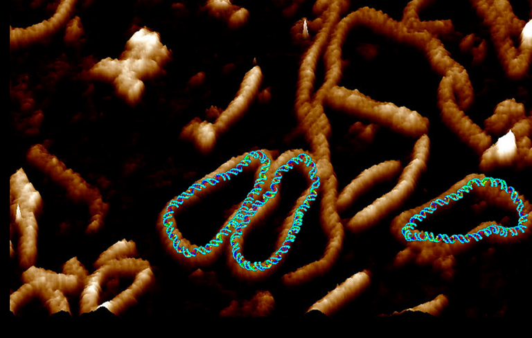
The footage is based on some of the highest resolution images of a single molecule of DNA ever captured, with DNA seen to “dance” in microscopy data recorded at the London Centre for Nanotechnology at UCL, and at the AFM manufacturer Bruker’s headquarters in Santa Barbara, California.
In the cell, DNA is arranged into highly-organised and supercoiled structures. Much is known about the molecular profile and function of DNA, however scientists are unclear about how this supercoiling affects the detailed double-helical structure of DNA, largely because of limitations in capturing images with a high enough resolution.
Previously scientists were only able to see DNA by using microscopes limited to taking static images. In the new study, published in Nature Communications, the research team combined advanced atomic force microscopy pioneered with supercomputer simulations to create videos of twisted molecules of DNA.
Further information
- Research paper: Base-pair resolution analysis of the effect of supercoiling on DNA flexibility and major groove recognition by triplex-forming oligonucleotides. Nature communications.
- UCL News: Video of ‘dancing DNA’ developed by researchers
- CRUK Drug – DNA Interactions Research Group, Professor John Hartley and Professor Daniel Hochhauser
- Kavit Main’s academic profile
- Dr Alice Pyne academic profile (University of Sheffield)
- London Centre for Nanotechnology
- UCL Physics & Astronomy
- UCL Mathematical & Physical Sciences
- Main image: Credit Dr Alice Pyne