Close
Close

Research for Non-Scientists
The human body is comprised of trillions of individual cells that must all work together for a common purpose. Key to the function of each of these cells are proteins, a type of molecule that act together as various molecular machines to replicate DNA (for gene inheritance), break down nutrients, fight off viruses, etc. Like any machine, a protein must be at the right place at the right time to perform its function properly. Clathrins (there two types in humans) are proteins that, amongst others, are responsible for transporting other proteins from one part of the cell to another, a process known as intracellular trafficking.
Specifically, the most well-studied role of clathrin is to move proteins from the cell surface to the inside of the cell, in a process known as endocytosis. Here, multiple molecules of clathrin weave together to assemble a basket-like coat on the inside surface (called a membrane) of the cell. This coat pulls the membrane to the inside and eventually breaks away, forming a ball of membrane, known as a vesicle, that is surrounded by a clathrin coat. Embedded within the membrane of these vesicles are cell-surface proteins, which are subsequently transported onwards to specific intracellular locations. One such application of this process is in the regulation of cell growth and division. Cells communicate with one another by releasing signalling molecules, which can provide cues to other cells, for example to stimulate these cells to divide. Once released, these signalling molecules can then be detected by receptors on other cells. The receptors can only detect the signalling molecule when they are located at the cell surface and in contact with the exterior environment of the cell. As such, by controlling how many receptors are at the cell surface through endocytosis, clathrin can control how the cells respond to exterior signals and hence when cells will divide.
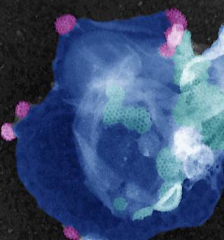
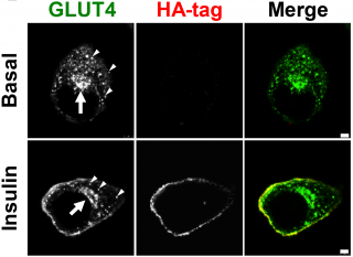
In addition to endocytosis, clathrins also partake in a wide variety of other membrane trafficking pathways, including the sorting of proteins into specialised intracellular regions of the cell and intracellular structures (called organelles—small organs). These specialised regions and organelles (bound by membranes) house bespoke cellular activities such as digestion and storage of transporters needed for uptake of cellular nutrients. The laboratory discovered a specialised form of clathrin responsible for packaging a sugar (glucose) transporter into a specific location within the cell. It is held here until blood sugar levels rise, and then (responding to insulin) is released to the cell surface to move the sugar from blood into muscle and fat cells. This process regulates blood sugar so that it is sufficiently high during fasting and reduces blood sugar after a meal when it would otherwise be too high. The latter is necessary because high levels of blood sugar can be poisonous to sensitive tissues. This specialised membrane traffic pathway for the glucose transporter is defective in type 2 diabetes, leading to the destructive effects of blood sugar accumulation.
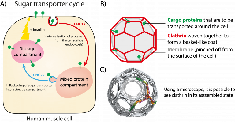
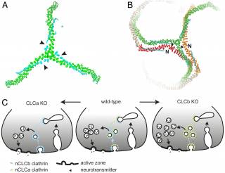
The two types of clathrin in humans are distinguished by their main structural protein, known as clathrin heavy chain (CHC). The historically well-studied CHC17 forms clathrin that regulates multiple pathways in all cells, and the less well-characterised CHC22 forms the specialised clathrin involved in glucose transport described above. The laboratory is currently investigating how these two CHCs operate and how they perform different trafficking functions in different cells. In addition, the CHCs are controlled by the binding of smaller regulatory proteins called clathrin light chains (CLCs). There are six different types of the CLC and the laboratory is studying how each of these fine-tunes the trafficking function of clathrin to meet the specific needs of different cell types.
In summary, clathrins act as a tiny molecular machine to capture proteins to move them around the inside of cells. This trafficking function plays a key role in a number of different cellular pathways ultimately being required for the development of organs composed of many cell types, the control of cell division, the regulation of nutrient metabolism, fighting infection and the proper functioning of the nervous system. Conversely, the malfunction of clathrin pathways can result in cancer, developmental defects, nervous system defects, Type 2 diabetes, and infectious disease. As a result, studying how clathrin functions on a molecular level provides a valuable understanding of how the body functions and also could provide promising targets for future pharmaceutical intervention.