Close
Close

Epithelial Physiology
Epithelia are sheets of tightly connected cells. They separate compartments within an organism or form the boundary between the organism and the external environment. They play vital physiological roles, from maintaining ideal homeostatic conditions in each compartment (ionic, pH, and volume balance), to exchanging gasses, to absorbing nutrients and excreting waste, to protecting the organism from dehydration and from threats present in the environment.
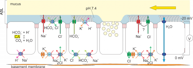
Carrying out these functions depends critically on epithelial cells maintaining “polarity”. In particular, the plasma membrane forms domains that have distinct lipid and protein composition on the cells’ apical (facing the external environment, e.g. gut lumen, alveolar sac, or cavity lumen) and basolateral (facing the internal environment and basement membrane) sides.
For instance, a layer of fluid is present on the apical surface of cells lining the airways. The ionic composition, pH, fluidity and volume of this Airway Surface Liquid (ASL) is carefully regulated by the action of distinct channel and transporter proteins that control transepithelial movement of water and solutes. These characteristics of the ASL are crucial for an innate defence mechanism, mucociliary clearance, that maintains the airways clean: particles and microorganisms get trapped in the mucus, and ciliary beat moves them to the pharynx to be swallowed.
Labs studying epithelial physiology in NPP focus mainly on channels and transporters of high clinical relevance, expressed in the airways, intestine and kidney.
Major themes include:
- Regulation of transepithelial glucose transport in kidney and intestine;
- Phosphate homeostasis;
- Pathophysiological consequences of mutations in KCNJ potassium channels;
- WNK pathway-controlled renal electrolyte and blood pressure homeostasis;
- Modelling and monitoring airway epithelia in health and in cystic fibrosis;
- Molecular pharmacology of the CFTR anion channel.
Research Labs


Recent Papers
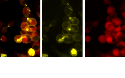
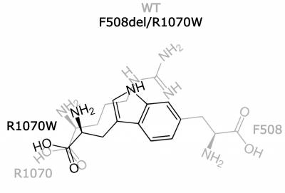
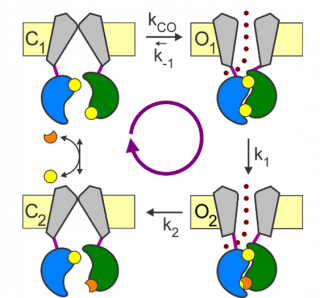
Can two wrongs make a right? F508del-CFTR ion channel rescue by second-site mutations in its transmembrane domains
Prins S, Corradi V, Sheppard DN, Tieleman DP, and Vergani P.
Why does the deletion of one phenylalanine (F508del) in CFTR cause so many problems to cystic fibrosis patients? Exploiting our newly developed CFTR-monitoring fluorescence assay we systematically introduce second-site mutations, in cis with F508del, in an intracellular loop known to be close in space to F508. Effective and poor "revertants" are then compared using molecular dynamics simulations to investigate the structural underpinnings of F508del defects.
Arginine-selective modulation of the lysosomal transporter PQLC2 through a gate-tuning mechanism
Leray, X., R. Conti, Y. Li, C. Debacker, F. Castelli, F. Fenaille, A. A. Zdebik, M. Pusch and B. Gasnier
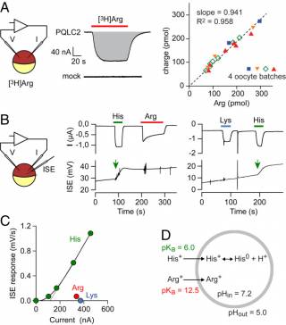
Lysosomes degrade excess or damaged cellular components and recycle their building blocks through membrane transporters. They also act as nutrient-sensing signaling hubs to coordinate cell responses. The membrane protein PQ-loop repeat-containing protein 2 (PQLC2; "picklock two") is implicated in both functions, as it exports cationic amino acids from lysosomes and serves as a receptor and amino acid sensor to recruit the C9orf72/SMCR8/WDR41 complex to lysosomes upon nutrient starvation. Its transport activity is essential for drug treatment of the rare disease cystinosis.
Defects in KCNJ16 Cause a Novel Tubulopathy with Hypokalemia, Salt Wasting, Disturbed Acid-Base Homeostasis, and Sensorineural Deafness
Schlingmann, Renigunta et al. 2021
The transepithelial transport of electrolytes, solutes, and water in the kidney is a well-orchestrated process involving numerous membrane transport systems. Basolateral potassium channels in tubular cells not only mediate potassium recycling for proper Na ⁺ ,K ⁺ -ATPase function but are also involved in potassium and pH sensing. Genetic defects in KCNJ10 cause EAST/SeSAME syndrome, characterized by renal salt wasting with hypokalemic alkalosis associated with epilepsy, ataxia, and sensorineural deafness.
Diet-induced iron deficiency in rats impacts small intestinal calcium and phosphate absorption
Evans O. Asowata, Oluwatobi Olusanya, Kaoutar Abaakil, Havovi Chichger, Surjit K. S. Srai, Robert J. Unwin, Joanne Marks
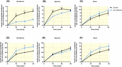
Iron deficiency appears to be a risk factor for the development of osteoporosis. Given that calcium and phosphate are key constituents of the bone mineral, hydroxyapatite, it is possible that the absorption of these minerals in the small intestine might be affected by iron deficiency. Therefore, the study aimed to systematically investigate the impact of iron deficiency on the cellular mechanisms of transcellular and paracellular calcium and phosphate transport in different regions of the rat small intestine. We demonstrated that iron deficiency alters calcium and phosphate transport in the duodenum and that this occurs via changes to the paracellular pathway and specific claudin proteins.
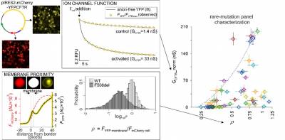
Fluorescence assay for simultaneous quantification of CFTR ion-channel function and plasma membrane proximity
Stella Prins, Emily Langron, Cato Hastings, Andra C. Stefan, Lewis D. Griffin, Paola Vergani
How well a protein does its job depends on it being in the right cellular location. CFTR is a channel protein, meant to be at the plasma membrane. Hundreds of different mutations in the CFTR gene cause cystic fibrosis. These affect, to a varying degree, how many channels reach the membrane (biogenesis), and how well they work (function).
We have developed an assay that simultaneously measures CFTR biogenesis and function. We used this tool for investigating CFTR mechanism, providing clues on how an approved drug works.
1,25(OH)2 vitamin D3 stimulates active phosphate transport but not paracellular phosphate absorption in mouse intestine
Nati Hernando, Eva Maria Pastor-Arroyo, Joanne Marks, Udo Schnitzbauer, Thomas Knöpfel, Matthias Bürki, Carla Bettoni, Carsten A. Wagner
It is well established that intestinal absorption of phosphate is stimulated by 1,25 dihydroxyvitamin D3 and that this involves enhanced NaPi-IIb expression and function. However, recent evidence suggests the presence of additional transcellular pathways for absorption and the likely dominance of a poorly characterised paracellular passive pathway. This study aimed to compare the effects of administration of 1,25 dihydroxyvitamin D3 in wild-type and intestinal-specific NaPi-IIb knockout mice and analysed the transcellular vs. paracellular routes of phosphate absorption. It was found that the hormone enhanced the active transcellular pathway for phosphate absorption, while the paracellular pathway was not affected. In NaPi-IIb knockout mice, 1,25 dihydroxyvitamin D3 failed to elicit a response, suggesting that the hormone specifically regulates NaPi-IIb expression.
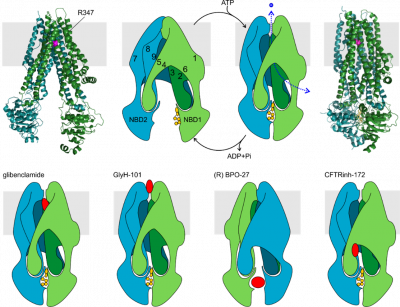
Strategies for cystic fibrosis transmembrane conductance regulator inhibition
Hugo R. de Jonge, Maria C. Ardelean, Marcel J. C. Bijvelds, Paola Vergani
While absence or dysfunction of CFTR causes cystic fibrosis, overactivation of CFTR, driven by microbial toxins (e.g. cholera toxin), underlies secretory diarrhoeas, a major cause of malnutrition and death of children under 5 years of age. Compounds that inhibit CFTR could improve emergency treatment of diarrhoeal disease. In this review we discuss how existing CFTR inhibitors function at the molecular and cellular level.
Although challenges remain, especially relating to the practical effectiveness of currently available CFTR inhibitors, we discuss how recent technological advances might help develop therapies to better address this important global health need.
A Nanosensor Toolbox for Rapid, Label-Free Measurement of Airway Surface Liquid and Epithelial Cell Function
Rositsa Ivanova, David C. H. Benton, Mustafa M. Munye, Sarinda Rangseesorranan, Stephen L. Hart, Guy W. J. Moss
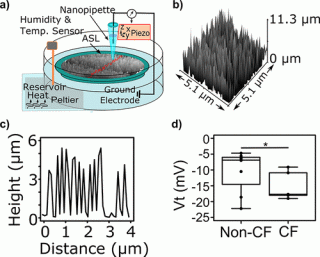
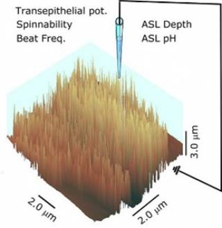
Breathing in air is essential to life but it can be a hazardous process as it brings with it bacteria, viruses and other unwanted particles. The body’s first line of defence against this sort of problem is a thin, fluid layer covering the upper airways called the airway surface liquid (ASL). The ASL contains sticky mucus designed to trap inhaled objects, which are then swept out of the lungs by the beating action of cilia (small finger-like projections from the cell surface). The composition and depth (~10 μm) of the ASL is tightly regulated by the body but in diseases such as cystic fibrosis, these control processes fail. In this paper we develop cutting edge nanotechnology that makes it possible, for the first time, to measure the depth of the airway surface liquid, the beating of cilia, the stickiness (rheology) of the mucus and many other important parameters without the need to add labelling molecules to the ASL that could distort the values being measured.
ACS Appl. Mater. Interfaces 2019, 11, 9, 8731-8739
Structure, Gating, and Regulation of the CFTR Anion Channel
László Csanády, Paola Vergani and David C. Gadsby
This comprehensive review describes the experiments that have revealed the molecular mechanisms underlying how anions permeate through CFTR, how its gates open and close, and how these movements are controlled.
But despite all we have learnt, there is still so much we do not understand!
The role of SLC34A2 in intestinal phosphate absorption and phosphate homeostasis
Joanne Marks
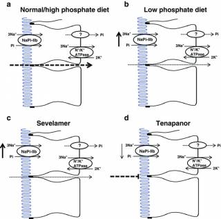
This invited review was published in a special issue of Pflügers Archiv - European Journal of Physiology, to coincide with the 25th anniversary of the publication of the cloning of NaPi-IIa (SLC34A1). The review summarises the most recent developments in our understanding of the role of the intestine in phosphate homeostasis. Evidence regarding the overall contribution of the transcellular and paracellular pathways for phosphate absorption is discussed, together with the clinical benefit of inhibiting these pathways for the treatment of hyperphosphatemia in chronic kidney disease.
Contact-Free Scanning and Imaging with the Scanning Ion Conductance Microscope
Samantha Del Linz, Eero Willman, Matthew Caldwell, David Klenerman, Anibal Fernández, Guy Moss
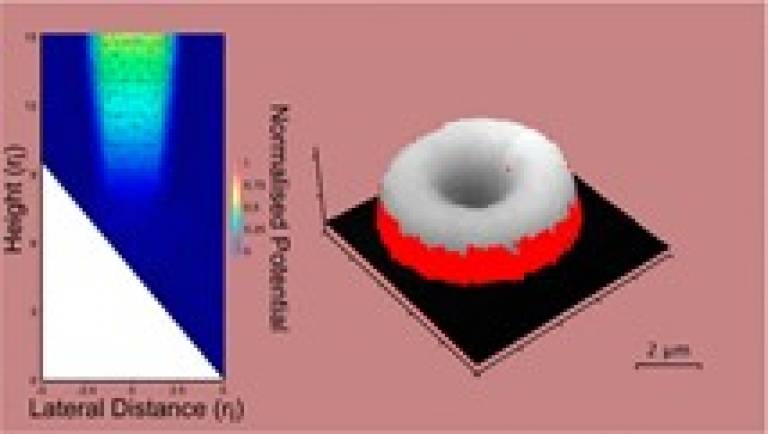
Anal. Chem. 2014, 86, 5, 2353-2360
Scanning ion conductance microscopy (SICM) is an imaging technique that allows researchers to obtain very high (nanometer) resolution topographical images of living cells. Unlike many other scanning probe techniques SICM can perform imaging without the need to contact the sample, which is a key advantage because touching a cell can trigger many changes. However, the conditions under which this type of scanning works, and what happens if these conditions are not met, is the subject of this paper. We used finite element modeling (FEM) to understand the contact-free mode of SICM and demonstrate the importance of getting the conditions right for accurate biological imaging.