 Close
Close

New publication and cover in the Biophysical Journal for Saric Lab
3 November 2020
Luke Davis wrote this short blog for the Biophysical Society communicating the science behind the Saric Lab's work for their new paper and cover in the Biophysical Journal. The title is inspired by the children's book ‘How the Zebra got its stripes.’
Type 1 collagen is the most commonly found protein in all vertebrates. It self-assembles into long string-like architectures called fibrils, which form the mechanical scaffold (matrix) of bone, skin, tendons, blood vessel walls, and other connective tissues. Collagen is also the major component of the network of fibrils that surrounds cells (the extracellular matrix), playing a key role in the determination of cell phenotype, cell adhesion, tissue regulation and signaling.
One of the defining features of type 1 collagen fibers is that they display a pattern of stripes when viewed under a microscope. This pattern is formed by a sequence of more dense and less dense fiber regions and is well preserved across the animal kingdom. What determines the striped fibrillar patterns in collagen? This question remained unsolved for decades. Using minimal models and computer simulations, we worked out the basic molecular features that govern the formation of striped fibrils. In brief, we find that fibril patterning is solely determined by the distribution of charges on the individual molecule, which leads to neat stacking or “stripes.”
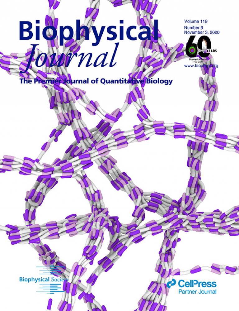
The cover image for the November 3 issue of Biophysical Journal is a computer simulation snapshot of the dynamic self-assembly of fibrillar structures using collagen-like molecules. The collagen-mimetic molecule contains a positively charged end (pink), a neutral central region (white), and a negatively charged end (purple). The charged ends are defined by a collection of charged units. The bundled network that is shown in the image displays regularly occurring gaps (white) and overlap areas (pink and purple), resulting from oppositely charged ends, attracted to each other through screened electrostatic interactions, falling in register. It is quite remarkable that a minimal model can replicate the occurrence of the stripe spacing similar to the one seen in real collagen fibrils, revealing a simple and robust process as to how collagen might have gotten its stripes.
Our study and this cover highlight the remarkable simplicity that is at the heart of robust biological assemblies such as collagen and the utility of so-called minimal coarse-grained modelling approaches. We hope our findings aid in the understanding of diseases associated with collagen fibrils and the design of artificial collagen mimicking architectures. Please see the animation here.
- Research paper in Biophysical Journal
- Blog for the Biophysical Society
- Dr Andela Saric's academic profile
- UCL Physics & Astronomy
- UCL Institute for the Physics of Living Systems