Prof Ferruccio Renzoni

Electromagnetic Imaging with Atomic Magnetometers
Our group has a unique expertise in electromagnetic induction imaging with atomic magnetometers. In 2013 we demonstrated the use atomic magnetometers in the magnetic induction tomography (MIT)-modality for the first time. In 2014 we realised the first demonstration of electromagnetic imaging with atomic magnetometers. This opened up a new realm for electromagnetic imaging, given the extreme sensitivity of atomic magnetometers at low frequency. Our breakthrough has generated considerable interest for a wide range of applications, from security and surveillance to industrial monitoring and medical imaging.
EMI with Atomic Magnetometers for Biomedical Applications
We are developing a novel quantum imaging platform based on ultra-sensitive atomic magnetometers (AMs) operating in the so-called magnetic induction tomography (MIT) modality, in unshielded environment. The superior performance of AMs and the contactless and non-invasive nature of MIT will provide 2D and potentially 3D maps of conductivity of biological tissue, thus filling the gap of current diagnostic tools. This would have a tremendous impact on the biomedical field, from fundamental research to diagnostics, in all cases where conductivity plays - or is expected to play - a relevant role.
People (current): Cameron Deans, Luca Marmugi, Ferruccio Renzoni
EMI with Atomic Magnetometers for Non-Destructive Evaluation
The capability of penetrating barriers and obstacles, and the possibility of non-invasively obtaining conductivity maps of object makes magnetic induction tomography very interesting for non-destructive evaluation (NDE) in industrial, corrosion, and health and usage monitoring. By pairing these capability with the performance of atomic magnetometers, we - in collaboration with NPL - are developing a novel quantum sensing platform, capable of assessing the structural integrity of components and materials, and their level of corrosion.
People (current): Cameron Deans, Luca Marmugi, Ferruccio Renzoni
Collaborations: Dr Witold Chalupzcak (National Physical Laboratory), Dr Rafal Gartman (National Physical Laboratory)
EMI with Atomic Magnetometers for Security and Surveillance
We are developing a multipurpose portable sensing and imaging platform, based on atomic magnetometers in magnetic induction tomography (MIT) configuration, for cargo and luggage screening, as well as remote detection and surveillance. Thanks to the sensitivity and tunability of atomic magnetometers, a novel instrument, capable of penetrating concealing barriers and to achieve long-range penetration in media, is envisaged. Furthermore, thanks to the inherent safety of MIT approach and the absence of ionising radiation, an extension of the application domains is expected, for example in the case of screening of delicate instrumentation, or when passengers, operators or stowaways might be involved.
Remote detection, localisation, and tracking of underwater non-magnetic objects is another crucial requirement. We have recently demonstrated the feasibility of an array of atomic magnetometers for active underwater surveillance.
People (current): Cameron Deans, Luca Marmugi, Ferruccio Renzoni


Magnetic Induction Tomography and Atomic Magnetometers
Our group was the first to demonstrate the feasibility of magnetic induction tomography (MIT) with atomic magnetometers (AMs): by combining these two technologies, a novel sensing and imaging platform was demonstrated. Whereas MIT had been already demonstrated in a number of fields, the limited performance of conventional magnetic field sensors and their limited tunability prevented broad practical applications, such as security - where penetration capability is paramount - or advanced biomedical imaging - where extreme sensitivity is a fundamental requirement. Thanks to MIT and the superior performance of AMs at room temperature, we are currently exploring novel paths, by pushing our setups to their limits.
MIT
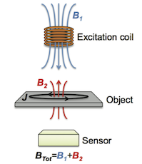
Magnetic induction tomography (MIT), also referred to as electromagnetically induction imaging (EMI), is a non-contact, non-invasive technique for mapping the dielectric properties (i.e. electric conductivity, permittivity and permeability) of an object of interest.
Its operation is based on the excitation of eddy currents in the object of interest by means of an AC magnetic field (primary B1). As a result of eddy currents, whose density is related to the dieletric characteristics of the medium they propagate through, a secondary AC magnetic field (B2), oscillating at the same frequency, is produced and opposes B1. The amplitude and the phase-lag of the secondary field in turn depend on the conductivity, permittivity and permeability of the sample. Therefore, by performing position-resolved measurements, maps of the dielectric properties (mostly conductivity) of an object can be realised.
However, MIT (or EMI) can be only as good as it is the sensor used for measuring the secondary field.
AM
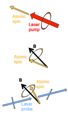
Atomic magnetometers (AMs) are quantum sensors based on laser interrogation of atomic spins, capable of state-of-the-art sensitivity (in some case, better than 10-15 T/Hz1/2), up to several orders of magnitude better than a standard pick-up coil of the same volume below 50 MHz. In addition, AMs can operate at room temperature (or at least without the need of cryogenics), in unshielded environments, and with low power consumption. Finally, they are well-suited for miniaturisation and - in certain configurations such as radio-frequency (RF) AMs - they can be tuned over broad bands. Therefore, AMs have the potential to be a 'game-changer' for MIT technology.
Broadly speaking, every AM is based on three steps, whose details can significantly differ from one implementation to another:
1) An atomic alkali vapour is spin-polarised via optical pumping;
2) Atomic spins precess in the magnetic field to be measured (Larmor precession);
3) Information on the spin motion and, consequently, on the characteristics of magnetic field to be measured are encoded in a probe laser beam, whose amplitude and polarisation are modulated by the Larmor precession.
References:
A. Wickenbrock et al., Appl. Phys. Lett. 103, 24, 243503 (2013)
DOI: 10.1063/1.4848196
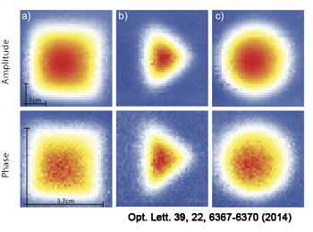
A. Wickenbrock et al., Opt. Lett. 39, 22, 6367-6370 (2014)
DOI: 10.1364/OL.39.006367
C. Deans et al., Appl. Phys. Lett. 108, 10, 103503 (2016)
DOI: 10.1063/1.4943659
C. Deans et al., Phys. Rev. Lett. 120, 033204 (2018)
DOI: 10.1103/PhysRevLett.120.033204
C. Deans et al., Rev. Sci. Instr. 89, 8, 083111 (2018)
DOI: 10.1063/1.5026769
EMI with Atomic Magnetometers for Biomedical Applications
Imaging is nowadays one of the most powerful tools for biomedicine and healthcare. However, no imaging technique is universal. None of the currently available technologies can provide maps of the dielectric properties, and in particular electric conductivity, of biological tissues. This can limit the possibilities of healthcare professionals, in particular for certain conditions such as atrial fibrillation.
To overcome these limitations, we propose to use atomic magnetometers (AMs) in the so-called magnetic induction tomography (MIT) configuration for non-invasively mapping the conductivity of tissues and organs.
Radio-frequency Rb Atomic Magnetometer
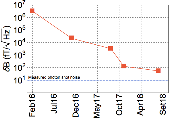
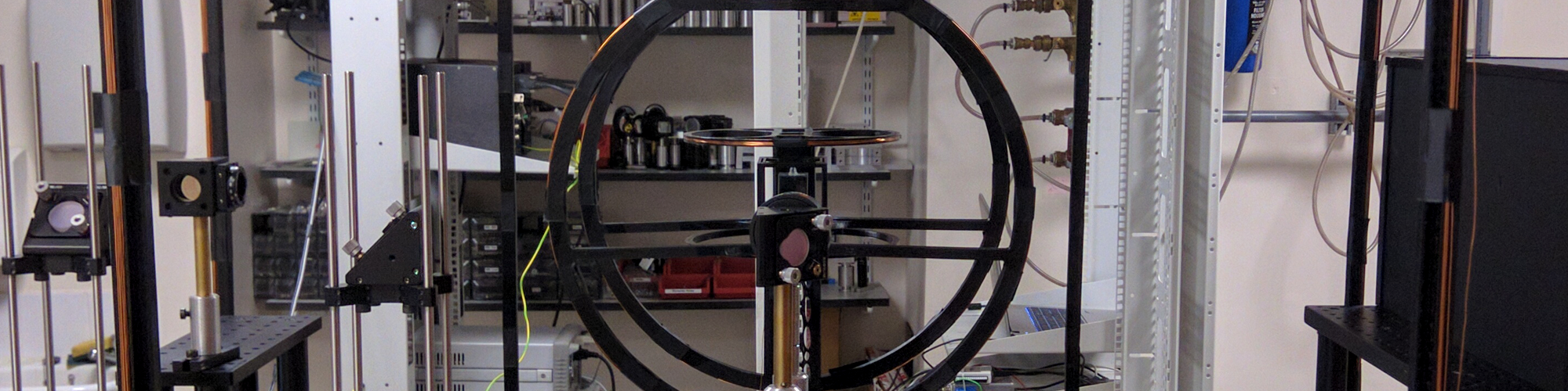
Our system is based on a radio-frequency (RF) atomic magnetometer, which can operate either with 85Rb or 87Rb. The core of the sensor is a quartz glass cell (side 25 mm) containing an isotopic mixture of Rb and 20 Torr of N2 to increase the atom/laser interaction. The laser light, tuned to the D2 line at 780 nm, is produced by a single DBR laser module. The beam is split in two branches, which act as a circularly polarised pump and as a linearly polarised probe. After optical pumping, an orthogonal AC field (driving field) coherently drives the AM at the desired frequency. The polarisation rotation due to Faraday rotation of the probe beam is then analysed by a balanced polarimeter and a lock-in amplifier. Active stabilisation of background stray fields allows operation in completely unshielded environment. In November 2017, our system reached a sensitivity of 130 fT/Hz1/2. After a number of upgrades, at the end of 2018, we reached 50 fT/Hz1/2 at 100 kHz, with single sensor and in unshielded environment. We also identified the limit of photon shot noise (see graph on the right). This makes it one of the most sensitive magnetometer of its kind currently deployed (December 2018).
EMI of Low-Conductivity Samples
For EMI, the driving field works also as the primary field of MIT: it induces eddy currents in the object of interest, which is moved with respect to the sensor by a computer-controlled XY translational stage. At each point, the sensor measures a total magnetic field (BTot), which contains also the secondary field produced by the eddy currents. By using phase-sensitive detection, information on the contribution of the secondary field are extracted. A map of the response of the target, consisting mostly of a conductivity map, is thus built in real-time by the computer-controlled data acquisition.
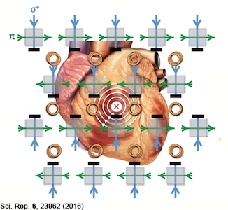
Application Study: Atrial Fibrillation
Information reported in this section is purely illustrative of our research project. It must not be used in any case as medical reference, nor does it substitute in any case for the advice of nurses, doctors, and other healthcare professionals.
Atrial fibrillation (AF) is a condition of the heart, which causes the normal electric activity of the heart to be altered and partially disrupted. As a consequence, the cardicac rhythm becomes irregular and faster during a crysis. Symptoms may vary, but usually include fatigue, shortness of breath, and sense of oppression on the chest. In the long term, however the anomalous activity of the atria (the top cavities of the heart) can cause severe secondary consequences, such as an increased risk of clotting and stroke, ventricular fibrillation, and sudden cardiac death. Because of this, AF is considered one of the main causes of morbidity and mortality in developed countries. It affects a significant fraction of the ageing population. Some estimates indicate that around one million persons above 65 years old are affected by AF in the UK only.
Despite these numbers, little is known about the causes of AF. It has been speculated that AF is caused by a change in conductivity of the heart tissue, and evidence supporting this hypothesis may have been recently found with the observation of rotors, circular patterns of electrical activity, in some patients referred for AF. The topic however is still widely debated. As a consequence, no direct and dedicated diagnostic tools are available for AF, unless a fibrillation crisis is on-going, and surgical treatment, involving scarring of the heart tissue has a non-ideal success rate.
We propose to investigate the causes of AF, and in particular to test the conductivity change hypothesis, by creating - for the first time - in vivo maps of the conductivity of the heart with our imaging system based on AMs. This will shed light on the underlying causes of AF and could potentially provide a new diagnostic tool and system for intra-operative guidance during surgical treatment of AF (Radio-Frequency Catheter Ablation, RFCA).
References:
L. Marmugi and F. Renzoni, Sci. Rep. 6, 23962 (2016)
DOI: 10.1038/srep23962
C. Deans et al., Appl. Phys. Lett. 108, 10, 103503 (2016)
DOI: 10.1063/1.4943659
C. Deans et al., Proc. SPIE, 9900, 9900F (2016)
DOI: 10.1117/12.2227538
C. Deans et al., Opt. Express 25, 15, 17911-17917 (2017)
DOI: 10.1364/OE.25.017911
C. Deans et al., Phys. Rev. Lett. 120, 033204 (2018)
DOI: 10.1103/PhysRevLett.120.033204
EMI with Atomic Magnetometers for Non-Destructive Evaluation
Evalution of fatigue or corrosion in industrial components or products is currently performed with different techniques and technologies depending on the context. Such surveys are usually invasive, e.g. requiring removal of covers and/or insulating layers, and often causes disruptions in the manufacturing process. This also further increases direct and indirect costs of such investigations. Therefore, tests are usually performed on a limited sample or only in a limited time frame. Consequently, timely and predictive maintenance, as well as detailed and thorough quality control during installation, are seldom possible. The costs of this for industry are in the range on billion of pounds per year.
We are therefore developing an imaging platform, suitable for operation in harsh environment, which would allow direct and non-invasive assessment of the structural integrity of metallic components, without any risk for operator and goods.
Proof-of-concept Demonstration
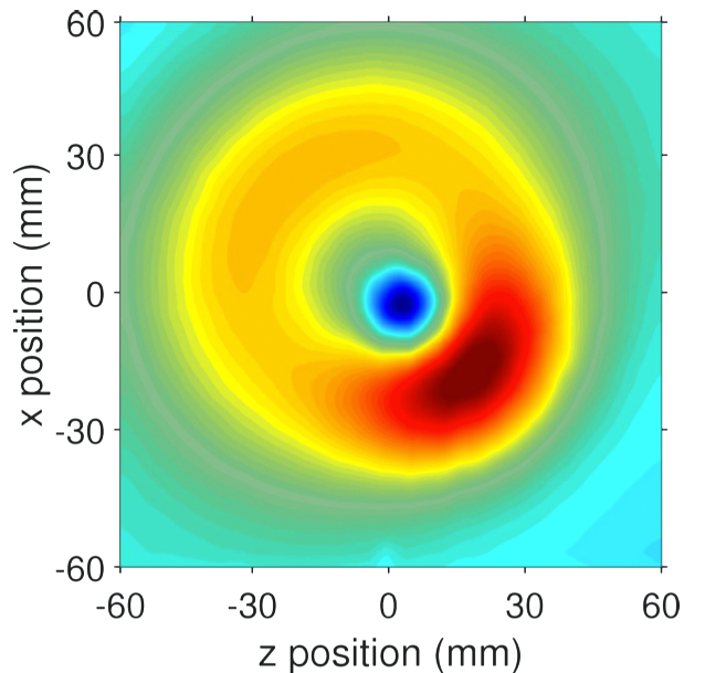
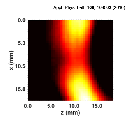
We have realised a series of proof-of-concept demonstrations, using a table-top radio-frequency (RF) 87Rb magnetometer operating in magnetic induction tomography (MIT) modality. For example, we have imaged and identified small cracks in conductive samples. As shown here on the right, we could dected sub-mm thick cut in an Al ring, despite the fact that this feature is smaller than the sensor and the excitation coil.
A second generation scanner is now being developed together with NPL for detection and imaging of corrosion.
Potential Applications
We envisage numerous applications for this instrument, ranging from quality monitoring during production and manufacturing, continuous remote monitoring of critical equipment, as well as on demand, spot-on surveying. All these functionalities would be achieved without any disruption of the manufacturing process, and without requiring removal of outer screens or protective layers. In fact, it is possible to penetrate thick conductive barriers by suitably tuning the operational frequency of our system.
We envisage a relevant impact on specific sectors of industry, as well as a significant reduction of maintenance costs, thanks to the possibiliy of timely intervention, before an actual damage occurs.
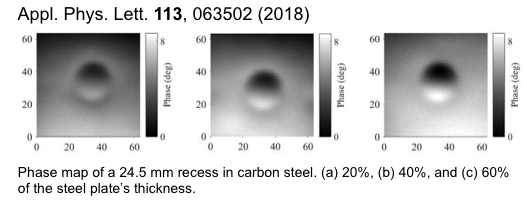
Corrosion Under Insulation Imaging with Atomic Magnetometers
Non-destructive inspection of structural steelwork, pipelines, and storage tanks is an open challenge. In particular, hidden corrosion is responsible for a large fraction of leaks, with severe consequences for production, costs, and the environment. We demonstrated detection and imaging of thinning of ferromagnetic carbon steelwork by using an atomic magnetometer operating in electromagnetic induction imaging modality, with a sensitivity of 0.1 mm. This approach is non-invasive, inherently safe, does not require disruption of production or the removal of insulating materials. These capabilities would provide tremendous advantages, allowing cost-effective monitoring and maintenance.
References:C. Deans et al., Appl. Phys. Lett. 108, 10, 103503 (2016)
DOI: 10.1063/1.4943659
C. Deans et al., Opt. Express 25, 15, 17911-17917 (2017)
DOI: 10.1364/OE.25.017911
P. Bevington et al., Appl. Phys. Lett. 113, 6, 063503 (2018)
DOI: 10.1063/1.5042033
EMI with Atomic Magnetometers for Security and Surveillance
Current instrumentation commonly deployed for security and surveillance suffer from a number of limitations, which reduce or hamper their applicability in critical cases. For example, standard metal detectors, based on coils as sensing elements, cannot penetrate to long ranges and are severely challenged with conductive barriers and cluttered environments. At the same time, X ray scanners are suitable only for short-range, and rely on the use of ionising radiation, which causes increase of costs, maintenance, and operation. Furthermore, it poses risks for operators, bystanders, drivers and passengers, as well as possible stowaways or animal cargos.
 We propose to overcome these limitations by using atomic magnetometers and magnetic induction tomography for imaging concealed targets in cargos, as well as performing remote detection, for example, of electrical motors' activity. A new instrument, with unprecedented capabilities, portable, cost-effective, safe, and potentially fully automated, can be thus envisaged.
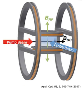
Portable RF Rb Atomic Magnetometer
One of the main goals of this research is to transfer the advantages of the AMs in EMI modality to a table-top and potentially portable device. This required - in a first phase - a complete redesign of the AM and its supporting instrumentation.
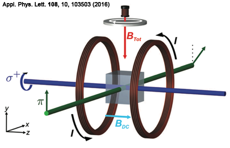
We have therefore realised a table-top platform, based on a RF 85Rb AM: a single laser source (an extended cavity system with intra-cavity interfence filter) polarises and interrogates Rb atoms contained in a quartz cube (25 mm side). The quartz cell contains also 20 Torr of N2 for increasing atom/laser interaction time. A dedicated 3D printed PLA support contains the vapour cell, and provides support for the DC and RF coils, the latter necessary for driving the magnetometer and generating the primary field for MIT operation.
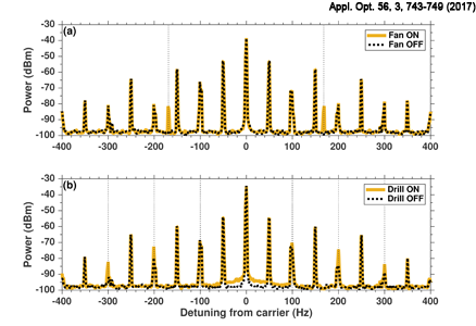
Remote Detection of Rotating Machinery
By relying on the coupling between our AM and the AC magnetic field generated by rotating machinery and electric motors, it is possible to remotely detect and monitor their activity. The AC magnetic signature of the rotating equipment appear, thanks to the non-linear mixing produced by the precessing spins in the atomic magnetometer, as sidebands in the power spectrum of the AM.Thanks to the sensitivity and tunability of the RF AM, we were able to detect AC and DC motors behind a wall and to identify their specific spectra.
These results demonstrate the potential for ultrasensitive devices for remote security and surveillance, as well as for industrial and usage monitoring (HUMS). In fact, by investigating the evolution of the collected spectra in time, timely maintenance can be scheduled, without the need of physically access the rotating equipment or interrupting its operation.
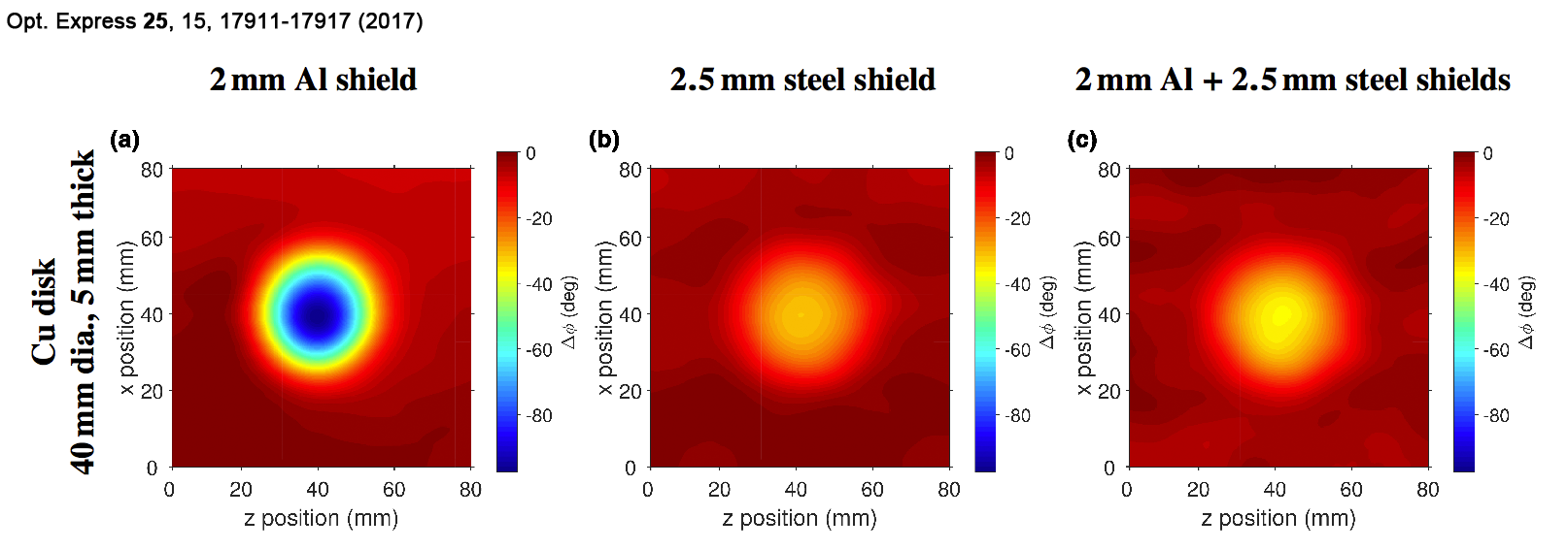
Safe EMI Screening Scanner
By using the sensitivity and tunability of RF AMs, we have demonstrated imaging of luggage and cargo screening in an inherently safe way, and without requiring access to the content of the container. In particular, by suitably changing the operational frequency of the system, it is possible to penetrate concealing barriers, even thick and conductive, to image the target underneath. Unlike other more conventional platforms, our system does not require dual imaging with background subtraction, or dual-frequency imaging: with a single scan and - in principle - without knowing the details of the concealing barrier, the target is detected and imaged.
This represents an important advance in the demonstration of our technology deployability: on the one hand, our scanner circumvents some of the limitations of conventional metal detectors, in particular in terms of tunability and sensitivity. On the other hand, our system will allow screening, without requiring access to the luggage or cargo, and without requiring the driver or other operator to leave the scene. This will increase the scanning throughput and improve the public's perception of the whole process.
Furthermore, by using an automatic stray field compensation system, the AM can operate in open environments also in presence of ferromagnetic material. In particular, we have demonstrated imaging through thick combinations of aluminium and ferromagnetic steel barriers, mimicking possible scenarios found during cargo or vehicle screening.
MIT Array for Underwater Detection and Surveillance
The detection and localisation of targets is possible using an array of atomic magnetometers performing MIT measurements. We used a 2x2 array to track underwater targets in real-time. The video below demonstrates the detection of multiple targets with live alarm triggering. In addition, we are able to track moving targets and suppress known background structures. Our results have applications from archaeological surveys to civil engineering. The active nature of the MIT approach is particularly relevant to underwater applications as it is immune to traditional magnetic countermeasures.
References:
C. Deans et al., Appl. Phys. Lett. 108, 10, 103503 (2016)
DOI: 10.1063/1.4943659
L. Marmugi et al., Appl. Opt. 56, 3, 743-749 (2017)
DOI: 10.1364/AO.56.000743
C. Deans et al., Opt. Express 25, 15, 17911-17917 (2017)
DOI: 10.1364/OE.25.017911
C. Deans et al., Phys. Rev. Lett. 120, 033204 (2018)
DOI: 10.1103/PhysRevLett.120.033204
C. Deans et al., Appl. Opt. 57, 10, 2346-2351 (2018)
DOI: 10.1364/AO.57.002346