How the brain gets wired for stereo vision
16 June 2011
Nerve cells that transmit light signals from the eye into the brain use a molecule best known for its role in blood vessel growth as a 'stepping stone' to help them reach the opposite brain hemisphere, according to research published in Neuron.

When light enters our eye, it passes through to the retina in the back of the eye, where it is converted to electrical signals that pass along nerve cells known as retinal ganglion cells. These ganglion cells follow a path into the brain, where they reach an area known as the optic chiasm. At this point, the path forks and the signal passes to both hemispheres of the brain - this is essential in order to create stereo vision, which allows us to see perspective.
However, until now, it has not been clear what guides the nerves to cross to the opposite side of the brain at the chiasm - why, for example, doesn't light from the right eye just pass to the right side of the brain rather than reaching to the left hemisphere?
The answer, according to Dr Lynda Erskine from the University of Aberdeen and Dr Christiana Ruhrberg, a Wellcome Trust New Investigator (UCL Institute of Ophthalmology)is that a molecule known as VEGF164, which ordinarily plays a part in the growth of blood vessels, acts as a stepping stone, allowing nerve cells carrying images from each eye to cross the chiasm and reach the opposite hemisphere.
The researchers developed a mouse model that allowed them to track how the ganglion cells are wired during fetal development. In mice lacking VEGF164, the axons of many retinal ganglion cell neurons do not cross to the opposite hemisphere.
"We found that the nerves from the eye grow along a path strewn with a small molecule that plays an important role in the growth and development of blood vessels," says Dr Ruhrberg. "This provides an interesting insight into the development of stereo vision, which relies on the balanced growth of axons from each eye into both brain hemispheres. We did not expect to find that nerve cells in the eye co-opt molecules essential for blood vessel growth to guide them to their destination."
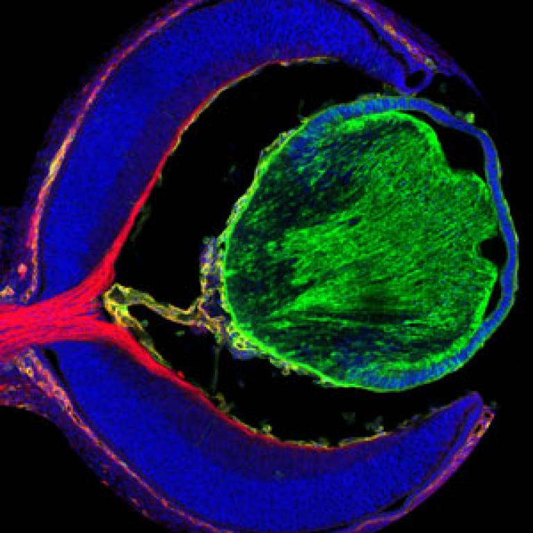
Image: Expression of the VEGF receptor NRP1
(red) on the axons of retinal ganglion cell neurons. The lens is green,
blood vessels are yellow and nuclei containing DNA are blue. (Credit:
Laura Denti.)
Links:
Wellcome Trust
UCL Institute of Ophthalmology
 Close
Close

