Close
Close

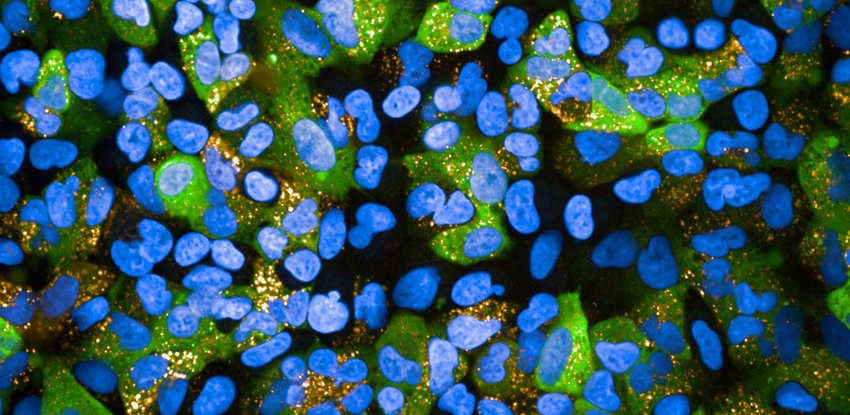
In cell biology, new approaches to imaging - including high-throughput imaging, super-resolution imaging and high-resolution datasets from 3D models or tissues - generate huge image data sets. In the past, such images could only be analysed visually, leading to a biased and incomplete set of possible interpretations.
The Bioinformatics Image Core (BIONIC) has trained bioinformaticians which link biology with mathematics, image processing, computer science and statistics to develop computational tools for image analysis. We use PerkinElmer Opera and Phenix high-content screening microscopes, ImageJ analysis software and the statistical program R with Bioconductor packages such as CellHTS2, in monolayer and 3D tissue culture.
Key methodologies include:
- Applied image segmentation
- Thresholding
- Morphological operations
- Filtering
- Feature extractions
- High-throughput statistical analysis
- Algorithm development