Meetings organized by the NEI and the FDA (13-14 March 2008 and 24 September 2010, Bethesda, MD, USA) have identified a clear and unmet need in glaucoma for methods to detect glaucomatous disease early in the disease process.
The current gold standard for the diagnosis of glaucoma is the measurement of visual fields.
However, it is estimated that up to 20-40% of RGCs are lost before field defects are detected using the gold standard detection method, and thus a potential 10-year delay in diagnosis exists currently.
Standard clinical tests are therefore clearly inadequate at detecting early glaucomatous visual functional deficits.
error message: 'NoneType' object has no attribute 'split'
Preclinical and clinical trials have explored a variety of compounds as possible neuroprotective agents in glaucoma with variable success to date.
Difficulties in translating from animal models to the clinic, in addition to the problems associated with clinical trial design in glaucoma, have contributed greatly to the paucity of neuroprotective agents that have reached Phase III clinical trials, despite the vast array of pre-clinical animal studies with positive results in the field.

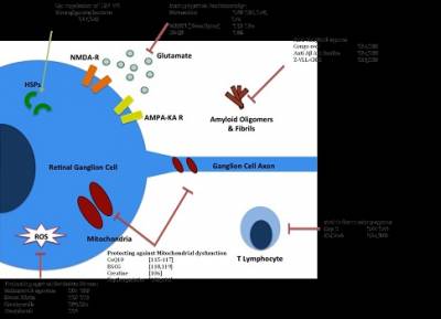
Potential neuroprotective agents in the treatment of glaucoma and their corresponding modes of action. Cop-1: Copolymer-1; CoQ10: Coenzyme Q10; DNQX: 6,7-Dinitroquinoxaline-2,3-dione; EGCG: Epigallocatechin galleate; Z-VLL-CHO: N-benzyloxycarbonyl-Val-Leu-leucine. From Expert Rev. Ophthalmol. Early online, 1-15 (2014).
We are using the DARC technology in in vitro and in vivo models of Glaucoma and RGC cell death to identify potential therapeutics for RGC apoptosis to meet this unmet need in the clinic, and as part of planned clinical trials, aim to assess its efficacy in diagnosing glaucoma patients up to 10 years earlier than the current gold standard.

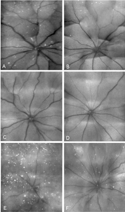
Visualization of RGC apoptosis using SLO imaging of in vivo glaucoma-related models. Staurosporine (SSP)-induced RGC apoptosis, appearing as white spots (A), was reduced by ifenprodil (0.6 nanomoles, B), MK801 (0.6 nanomoles, C), and LY354740 (20 nanomoles, D) at 2 hours after treatment. Similarly, RGC apoptosis at 3 weeks in a rat OHT model (E) was significantly reduced by treatment with a combined low dose of MK801/ LY354740 (0.06/20 nanomoles, F). From Invest. Ophthalmol. Vis. Sci.February 2006 vol. 47 no. 2 626-633
 Close
Close

