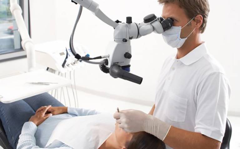
The dental operating microscope in daily practice
1 November 2016

By Greg Finn
Do you ever have a problem deciding whether there is a crack in a tooth? Can you be sure that you have removed the caries hiding in the annoying shadow in a distal box form on an upper molar? If so, are you sure there isn’t an exposed pulp horn? Do you have difficulty choosing which driver fits into that loose abutment screw the patient has arrived with?
In 1997 I asked one of my endodontic gurus, Richard Kahan, what piece of equipment I should invest in first? He is an equipment man through-and-through, but number one on his list at that time, before rotary preparation instruments, before warm vertical obturation systems, before endosonics, before digital radiography was the dental operating microscope.
Predictably, the microscope transformed my ability to identify canal openings, to differentiate sclerosed dentine and to see cracks. What surprised me was just how useful it was when preparing a cavity or checking the fit of a crown. I rapidly discovered that the scope became an indispensable part of everyday dental practice.
I am able to sit upright instead of spending my days bent over my patients. Over the same years, I have seen many of my contemporaries develop back, neck, wrist and shoulder problems. I am sure much of this can be tracked back to their habitual dental posture. According to Bethany Valachi (2008), 30% of dentists who retire early do so because of a musculoskeletal disorder. Whilst one in eight people in the general population suffer from low back problems annually, the figure is one in two amongst dentists and hygienists. Ergonomics is clearly a major issue for dental professionals. The operating microscope allows me to work with my spine upright, my hands at around elbow height and my eyes looking straight ahead and focused on infinity, their most relaxed state.
I also understand that the average 50-year-old requires twice the amount of light to achieve the same acuity as they had when they were 25. The illumination I get from the microscope allows me to see all the detail I could when I was younger. I once met McSpadden the endodontist who mid-70s book showed his operating microscope. At the time he told me “the hand can do what the eye can see.” This is something I have found to be true. The average person needs to wear reading glasses once they reach their mid-forties. However, with appropriate magnification and illumination a parallel decline in our manual dexterity does not have to accompany aging.
It is my hope that the extra years I will be able to work, should I wish to, plus the years I hope I to be free of chronic back pain in my retirement, hugely outweigh any financial investment. For me, I feel that purchasing the operating microscope has been one of my better decisions.
However, you don’t need to commit to buying a microscope to see whether it suits you. The UCL Eastman offers day courses where you can use operating microscopes in both restorative and endodontic settings:
Dental Microscopy: Endodontics
A one-day course focusing on the dental operating microscope in endodontics. Book now for December.
Dental Microscopy: Restorative Dentistry
A one-day course focusing on the dental operating microscope in restorative dentistry. Book now for December.
 Close
Close


