 Close
Close

As part of the Deep Imaging Mummy Cases project, seven different imaging modalities were used, each with a promise of its own. They are described here.
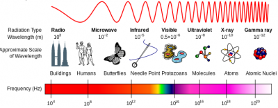
The electromagnetic spectrum
We used a range of different imaging modalities in this project, using light, x-rays and terahertz radiation. All these are electromagnetic waves, so we'll start with a reminder of what those are.
Different types of electromagnetic radiation are best distinguished by their wavelength. Optical radiation is radiation which we can see (visible), plus radiation which is slightly beyond the visible (near ultraviolet and near infrared). This has wavelengths of a bit less than a micrometre - smaller than a red blood cell. We perceive the wavelength of visible light as different colours, which are characteristic of inks and dyes.
The Electromagnetic Spectrum
Terahertz (THz) radiation has longer wavelengths than optical, up to about a millimetre. Certain materials (e.g. clothing and paper) are largely transparent to THz whereas others (e.g. water) absorb THz radiation heavily. We expect, therefore, that THz radiation will give different, and hopefully complementary information to optical radiation.
We are all familiar with the medical applications of x-rays. X-rays have very short wavelengths, around a nanometre, which is similar in size to small molecules. X-rays penetrate most materials and can be used in many ways, such as to image the density of a material, or to determine its elemental composition.
Other forms of electromagnetic radiation such as microwaves and radiowaves have wavelengths which are much longer than the objects we are interested in. They don't interact with the objects in any helpful way so we don't use them.
Multispectral imaging
Different dyes and inks appear to have different colours. Our eyes, and the cameras which most of us are familiar with, can detect three different colours: red, green and blue. To analyse the colour of an object with more precision than this, we can turn to multispectral imaging. Extending the analogy with vision, we could choose to illuminate with white light and, instead of detecting three colours, we can use a range of filters to identify different colours. We used this approach in a previous project. However, we now use an improved system which illuminates with fixed, discrete wavelengths (thereby reducing the total intensity of the illumination) and then detect with a high resolution camera potentially with filters (to identify fluorescent light).
Multispectral imaging has been shown to highlight otherwise illegible details in items as diverse as the Archmides Palimpsest, the Syriac Galen Palimpsest, and Livingstone's diaries. It can reveal information about the condition and composition of a document. It is safe, non-destructive and is developing a reputation as the first choice imaging modality for fragile manuscripts.
Our imaging system was provided by RB Toth Associates and consists of a 60 megapixel Phase One IQ260 achromatic camera , with a filter wheel with five long-pass filters, and computer-controlled lighting with 12 different wavelengths from ultraviolet to near infrared supplied by Dr Bill Christens-Barry of Equipoise Imaging LLC.
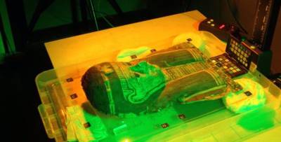
Multispectral Imaging in progress at the UCL Digitisation Suite
Optical Coherence Tomography
Optical Coherence Tomography (OCT) uses similar wavelengths to multispectral imaging, but works at a much smaller scale. It is best known as a medical imaging technique which is commonly used to image the eye and the skin. The system is arranged so that light travels a similar distance along two paths; one a reference arm and the other interrogating the sample. As light penetrates the sample and reflects back off different layers, the distance it travels changes. This change in distance is measured very precisely using a technique called interferometry. It can image to depths of around a millimetre and give micrometer resolution, so it is well suited to imaging layered structures.
OCT's use in imaging historical manuscripts is still experimental but we felt that its advantages, of non-contact depth-resolved imaging of pigments suggested that it could be a valuable addition to our imaging options.
Terahertz imaging
Terahertz (THz) imaging uses electromagnetic waves which are longer than visible waves. They can penetrate further through dry, insulating samples than visible waves but are heavily attenuated by water and metals. In dry samples such as our mummy cartonnages, we expect to be able to penetrate by 1cm or more, potentially allowing us to see through plaster, cloth or other coverings. Like OCT, its application to imaging manuscripts is experimental, though some early results in cultural heritage including manuscripts have been reported.
Terahertz imaging can be carried our in different modes. Imaging can provide information over an extended area, and spectroscopy can provide more detailed data about the constituent chemicals making up a sample.
X-ray fluorescence
X-ray fluorescence is a well-established technique which analyses secondary x-ray emitted from a sample whilst it is irradiated. The secondary x-rays are characteristic of a particular element, so analsis of the XRF spectrum can determine the composition of a sample. XRF is particularly valuable when identifying heavy metals within a sample. Under good conditions, the sensitivity can be as high as a few parts per million.
The highest quality x-ray beams are only available at synchrotrons, but that requires the sample to be moved. Despite that, synchrotron imaging of heritage objects is becoming a standard tool. Some objects, however, cannot be moved, so portable hand-help XRF devices have been developed for use outside the laboratory.
We used a Olympus Delta Premium handheld x-ray fluorescence device which we have been kindly loaned by UCL Institute of Archaeology. This is a flexible, portable device which is used in archaeology and in art and heritage applications. We expect that it will be able to identify the presence of ink beneath the surface of the object and give information about its elemental composition.
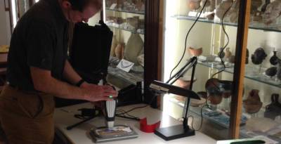
X-ray fluorescence system in use in the Petrie Museum, UCL
Phase contrast x-ray
We are used to medical x-ray images which detect x-rays which have passed straight through the body and made a "shadow" showing areas which attenuated the x-rays most strongly. However, the x-rays which pass through the sample develop a phase shift, depending on the material which they passed through. This phase shift can be measured and can provide contrast to changes which are not visible to standard medical x-rays.
X-ray phase contrast imaging was largely developed for medical imaging at synchrotons, but has been used to image a wide range of heritage objects. Recently, phase contrast x-ray was used to image the Herculaneum scrolls at the European Synchrotron at Grenoble and showed some evidence of text. Our laboratory at UCL has developed an approach for laboratory-based phase contrast x-ray [see movie].
X-ray microCT
X-ray CT is another imaging modality which was developed for medical applications and has famously been directly translated into heritage science by imaging intact mummies. X-ray micro CT works on a much smaller scale and is used to image objects like small animals, medical samples, electronics and materials with resolution of as small as a few micrometres.
It is used in palaeontology and analysis of artwork especially sculptures. We used it to determine the internal structure of the mummy cartonnage fragments to assist the interpretation of other imaging techniques.
Findings
Our comparison and approach showed that no current single imaging technique can identify both iron and carbon based inks at depths within cartonnage. If we are to detect and ultimately read text within cartonnage, a multimodal imaging approach is required, but this will necessarily be limited by cost, access to imaging systems and the portability of both the system and the cartonnage.
We are currently working on publications that will elucidate our approach and detailed findings, and we will share these here when published.